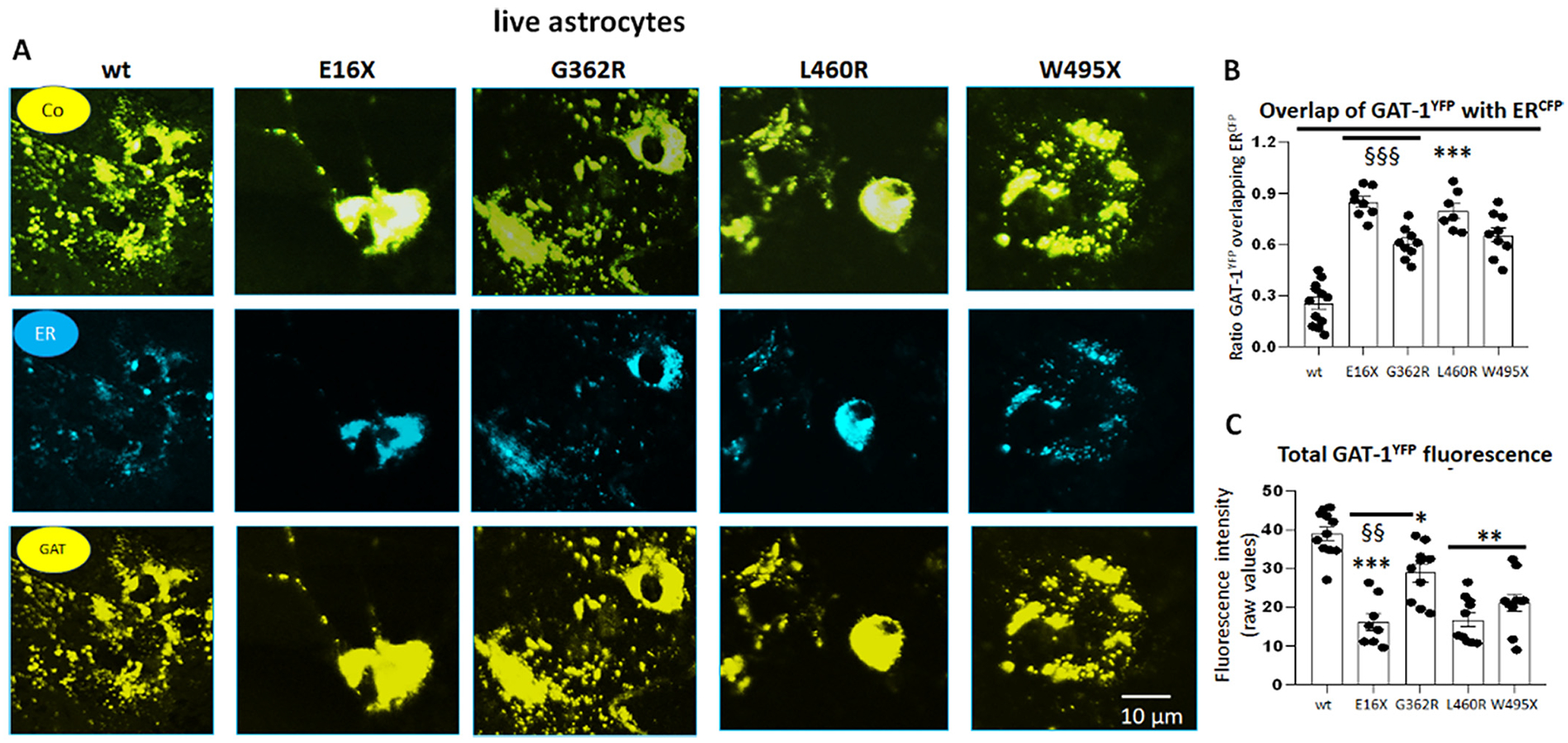
Fig. 5.

The mutant GAT-1 had reduced total protein expression but increased localization inside the endoplasmic reticulum in astrocytes.
A. Mouse cortical astrocytes under passage 2 were transfected with wildtype GAT-1YFP or the variant GAT-1 (E16X)YFP, GAT-1 (G362R)YFP, GAT-1 (L460R)YFP and GAT-1 (W495X)YFP with the pECFP-ER marker (ERCFP) at a 1:1 ratio (1 μg:1 μg cDNAs) for 48 h. Live cells were examined under a confocal microscopy with excitation at 458 nm for CFP and 514 nm for YFP. All images were single confocal sections averaged from 8 times to reduce noise, except when otherwise specified. “Co” stands for overlay, “ER” stands for endoplasmic reticulum marker (ERCFP) marker, and “GAT” stands for GAT-1YFP. (B). The GAT-1YFP fluorescence overlapping with ERCFP fluorescence was quantified by Metamorph with colocalization percentage. (C). Total fluorescence intensity (raw values) over the area was measured for each whole field and the raw mean values were plotted. In B and C, ***p < 0.001 mutant vs. wt, §§ P < 0.01, §§§ < 0.001 vs G362R. N = 7–8 representative fields from 4 different transfections. One-way analysis of variance (ANOVA) and Newman-Keuls test was used to determine significance compared to the wt condition and between mutations. Values were expressed as mean ± S.E.M.
