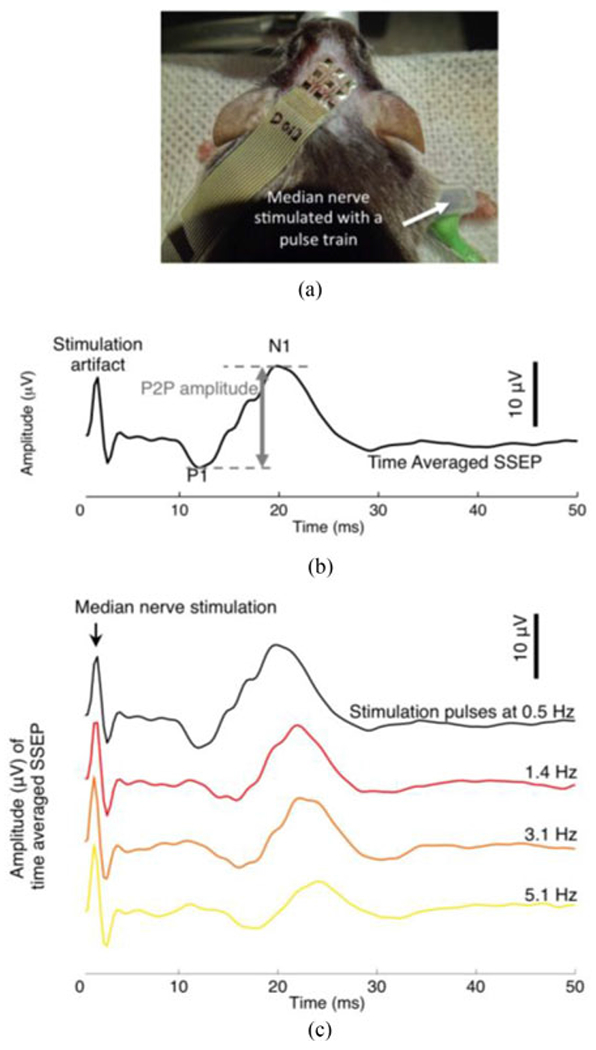
Fig. 3.

(a) A flexible electrode array positioned on the scalp of an anesthetized mouse during SSEP recording. (b) A time-averaged SSEP in response to electrical stimulation of the median nerve. The stimulation consisted of a pulse train of 0.2 ms biphasic pulses repeated at 0.5 Hz. Median nerve stimulation elicited prominent SSEP peaks, in this particular case, at ~13 ms (positive polarity, P1) and ~20 ms (negative polarity, N1). The SSEP shown is the average of 200 individual evoked trials. (c) Time-averaged SSEPs in response to median nerve electrical stimulation pulses delivered at a pulse frequency of 0.5 Hz, 1.4 Hz, 3.1 Hz and 5.1 Hz. Each averaged SSEP trace is the average of 200 individual evoked trials.
