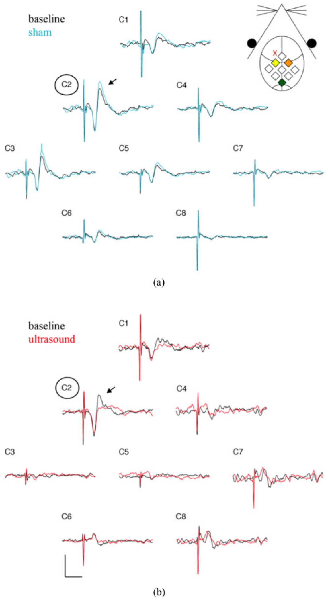
Fig. 7.

SSEP recordings from all recording electrode contacts. (a) Baseline (black) and sham (blue) SSEP recordings from each of the recording contacts (C1-C8). A schematic of channel position with respect the mouse anatomy is shown in the inset. Channel C2 (yellow diamond) is positioned over the somatosensory cortex contralateral to the stimulated median nerve, while channel C4 (orange diamond) is positioned over the ipsilateral somatosensory cortex. Channel C2 records the largest SSEP amplitude (arrow). Sham stimulation does not alter the amplitude for any channel. (b) Baseline (black) and ultrasound perturbed (red) SSEP recordings for each channel. Ultrasound application, located at the red “X” in the inset, reduces the amplitude of the recorded SSEP in contralateral somatosensory cortex (C2). Scale bar is 10 μV and 20 ms.
