FIG 3.
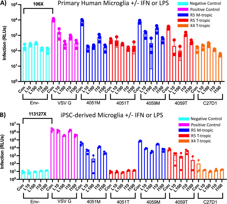
LPS and IFN restrict HIV-1 infection in primary and iPSC-derived microglia, but cell models are not equally permissive. (A and B) Primary human microglia (A) and human iPSC-derived microglia (B) were treated with 10 ng/mL (L10) and 100 ng/mL lipopolysaccharide (LPS) or interferon alpha (IFN-α) for 24 h and then infected with HIV-1 luciferase reporter viruses pseudotyped with patient-derived R5 macrophage-tropic (blue), R5 T cell-tropic (red), or X4 T cell-tropic envelope proteins. Untreated control groups are labeled “Con” within each Env group. Negative-control viruses (cyan) lacked an envelope protein, whereas the positive-control viruses (pink) were pseudotyped with vesicular stomatitis virus G protein, which is capable of infecting a wide range of mammalian cells. The average magnitude difference between the positive and negative control was 106× for primary microglia and 113,127× for iPSC-derived microglia. Infection was measured in relative light units (RLUs) via luminometer. N = 3 technical replicates.

