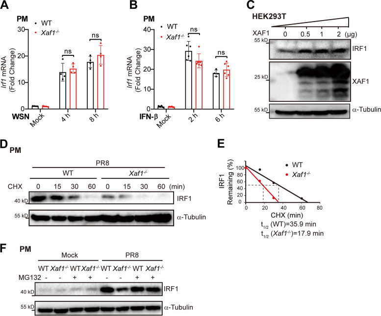
FIG 8.
XAF1 inhibits the proteasome degradation of IRF1. (A) qRT-PCR analysis of Irf1 mRNA level in the WT and Xaf1−/− PMs infected with WSN (MOI 0.1) for the indicated time points. (B) qRT-PCR analysis of Irf1 mRNA level in the WT and Xaf1−/− PMs treated with IFN-β (500 U/mL) for the indicated time points. (C) The XAF1 plasmid (0, 0.5, 1, or 2 μg) was transfected into HEK293T cells in a 6-well plate. After 24 h, total cell lysates were subjected to immunoblot analysis of indicated proteins. (D and E) The WT and Xaf1−/− PMs were infected with PR8 (MOI 0.1) for 8 h. After CHX (100 μg/mL) treatment, cells were harvested at the indicated time points, and cell lysates were subjected to immunoblot analysis (D); and the optical density of IRF1 protein bands was analyzed with ImageJ software (E). (F) The WT and Xaf1−/− PMs were infected with PR8 (MOI 0.1) for 6 h, followed by MG132 (10 μM) treatment for 6 h. Cell lysates were subjected to immunoblot analysis. (A and B) Data from three independent experiments are presented as mean ± SD; ns, no significant difference by unpaired Student's t test. (C–F) Data are representative results of three independent experiments.

