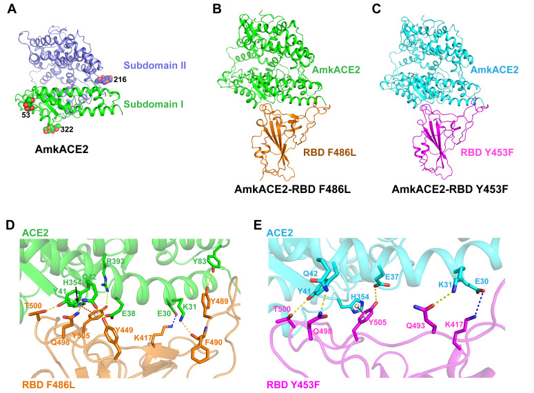
FIG 3.
Complex structure of AmkACE2 bound to RBD F486L or RBD Y453F. (A) Cartoon structural representation of AmkACE2. Subdomain I (S19-Q102, N290-N397, and P415-E430) and subdomain II (S103-P289, E398-T414, and D431-D615) (57) are colored in green and blue, respectively. The glycan N-linked sites are labeled. (B and C) Cartoon structural representations of AmkACE2-RBD F486L (B) and AmkACE2-RBD Y453F (C). RBD F486L and RBD Y453F are colored in orange and magenta, respectively. (D and E) Detailed interactions between AmkACE2 and RBD F486L (D) or RBD Y453F (E). The key contact residues are shown as stick structures and labeled. Hydrogen bond interactions were analyzed at a cutoff of 3.5 Å and are colored in yellow. The salt bridge is colored in blue. The π-π stacking interaction is colored in red.

