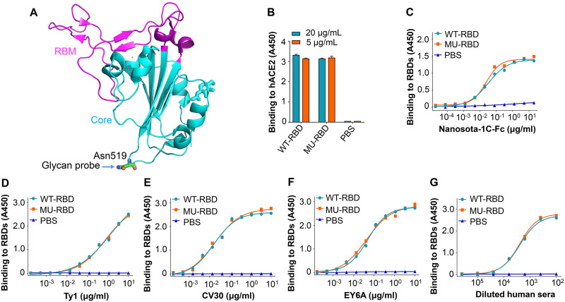
FIG 1.
Introduction of glycan probe and characterization of glycosylated mutant SARS-CoV-2 RBD protein. (A) Crystal structure of SARS-CoV-2 RBD (PDB access code: 6M0J). The core structure is colored in cyan, and the receptor-binding motif (RBM) in magenta. Mutated residue (Asn519) is shown where an N-linked glycan probe was introduced. (B) Receptor-binding affinity of mutant receptor-binding domain (RBD) (MU-RBD) subunit vaccine. An enyzme-linked immunosorbent assay (ELISA) was carried out to assess the binding of MU-RBD protein to soluble human angiotensin-converting enzyme 2 (hACE2) protein. Prototypic wild-type (WT)-RBD protein was included as comparison. (C to G) Antibody-binding affinity of MU-RBD subunit vaccine. ELISA was carried out to detect the binding of MU-RBD protein to SARS-CoV-2 RBD-specific neutralizing nanobodies Nanosota-1C-Fc (C) and Ty1 (D), neutralizing MAbs CV30 (E) and EY6A (F), and neutralizing human sera (G). WT-RBD was used as comparison. Data (panels B to G) are presented as mean ± standard error of the mean (SEM) of quadruple wells. The experiments were repeated twice, resulting in similar results.

