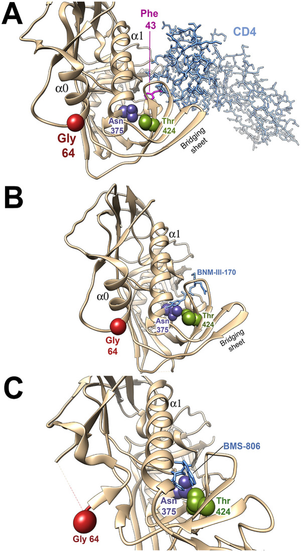
FIG 3.

Location of amino acid residues implicated in virus resistance in Env structural models. The gp120 amino acid residues implicated in resistance to the inhibitors are shown on structures of HIV-1 Env-inhibitor complexes. In all structures, the resistance-associated changes have been introduced by the swapaa function in Chimera (115). (A) The HIV-1 gp120 core with N/C termini (tan ribbon) complexed with two-domain CD4 (light blue sticks) (PDB: 3JWO) is shown (87). Phe 43 of CD4 is colored magenta. (B) The HIV-1 gp120 core with N/C termini (tan ribbon) complexed with BNM-III-170 (light blue sticks) (PDB: 5F4P) is shown (87). (C) The HIV-1 sgp140 SOSIP.664 trimer (tan ribbon) complexed with BMS-806 (light blue sticks) (PDB: 6MTJ) is shown (82). The Envs are oriented with the viral membrane at the top of the figure and the target cell membrane at the bottom. The α0 and α1 helices of gp120 are shown. Note that in panel C, the α0 helix is not present. Part of the gp120 layer 1 loop between residues 59 and 64 is disordered in 6MTJ, and therefore the location of Gly 64 is an approximation.
