FIG 6.
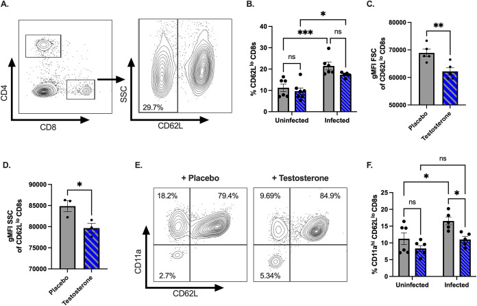
Testosterone dampens the activation of CD8+ T cells following oral CVB3 infection. (A) Representative flow cytometry plot of the gating strategy by which to identify CD62Llo CD8+ T cells. (B) The frequency of CD62Llo CD8+ T cells in the spleen of testosterone-treated (blue with diagonal lines) and testosterone-depleted (gray) male mice. *, P < 0.05, **, P < 0.01, ***, P < 0.001, ns: not significant; two-way ANOVA. (C) The geometric mean fluorescence intensity (gMFI) forward scatter (FSC) of CD62Llo CD8+ T cells **, P < 0.01; unpaired t test. (D) Representative of the gMFI side scatter (SSC) of CD62Llo CD8+ T cells from two independent experiments. *, P < 0.05; unpaired t test. (E) Representative flow cytometry plots of the expression of CD11a and CD62L CD8+ T cells in the spleen of testosterone-treated (blue with diagonal lines) and testosterone-depleted (gray) male mice. (F) The frequency of CD11ahiCD62Llo CD8+ T cells in the spleen following CVB3 infection. *, P < 0.05; two-way ANOVA. All data are from two independent experiments with n = 5 to 6 mice per group and are shown as mean ± SEM.

