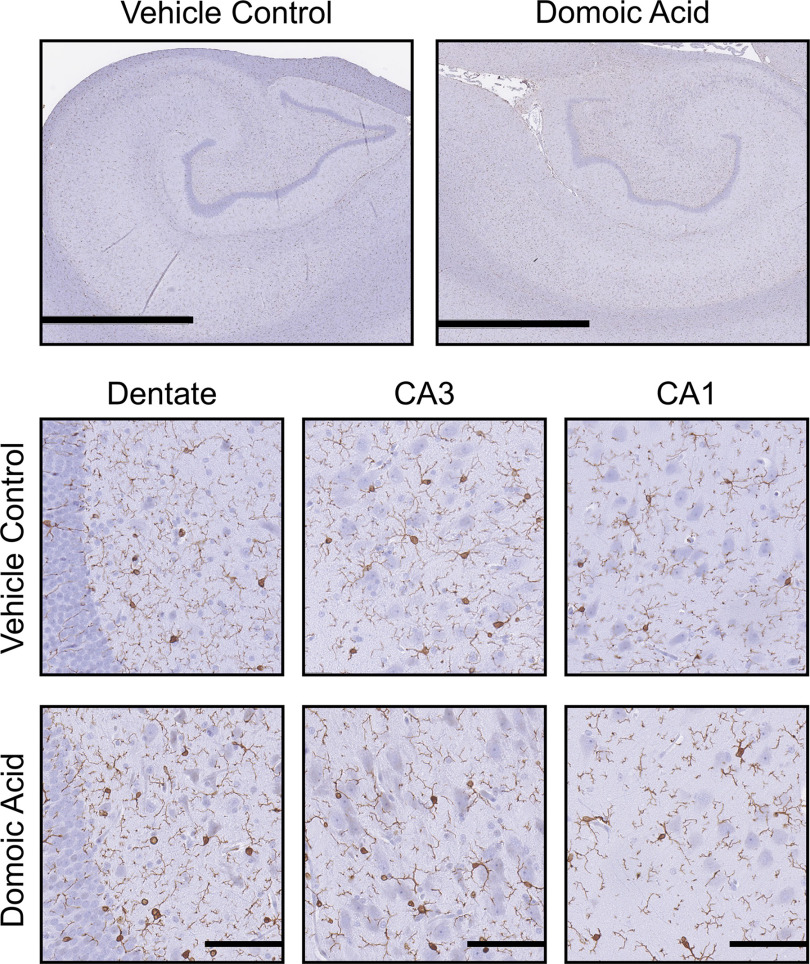
Figure 5.
Hippocampal Iba-1 immunoreactivity. Representative images of immunostaining of 10% formalin-fixed, paraffin-embedded, sections for (1:2,000; Wako Chemicals) microglia in the hippocampus of female Macaca fascicularis following prolonged exposed to domoic acid () or vehicle (5% sucrose). Images represent the hippocampus () and specific hippocampal regions, including the dentate gyrus (scale bars: 200 and ) and the CA3 and CA1 pyramidal cell layers (). Immunoreactivity was visualized with Vectastain Elite and shows as darker process-bearing cells within the image. Sections were counterstained with cresyl violet (CV). Note: CA, cornu ammonis area; , ionized calcium binding adaptor molecule 1.

