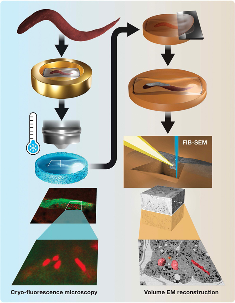
FIG. 1.
Schematic for cryo-LM/FIB-SEM workflow. Left panel: C. elegans worms are enclosed in cellulose capillaries and high-pressure frozen in planchettes (top). Dividing embryos in the intact worm are imaged in situ by cryo-fluorescence microscopy, revealing targeted and unexpected or rare events (bottom, green, SP12::GFP, red, H2B::mCherry). Right panel: Sectioning of a freeze-substituted sample reveals a cross section of the worm (top), which is used to position the FIB-SEM trench and image acquisition. The resulting nanoscale EM image volume is examined, and correlatively identified features of interest are reconstructed in 3-D (bottom).

