
Fig. 3.
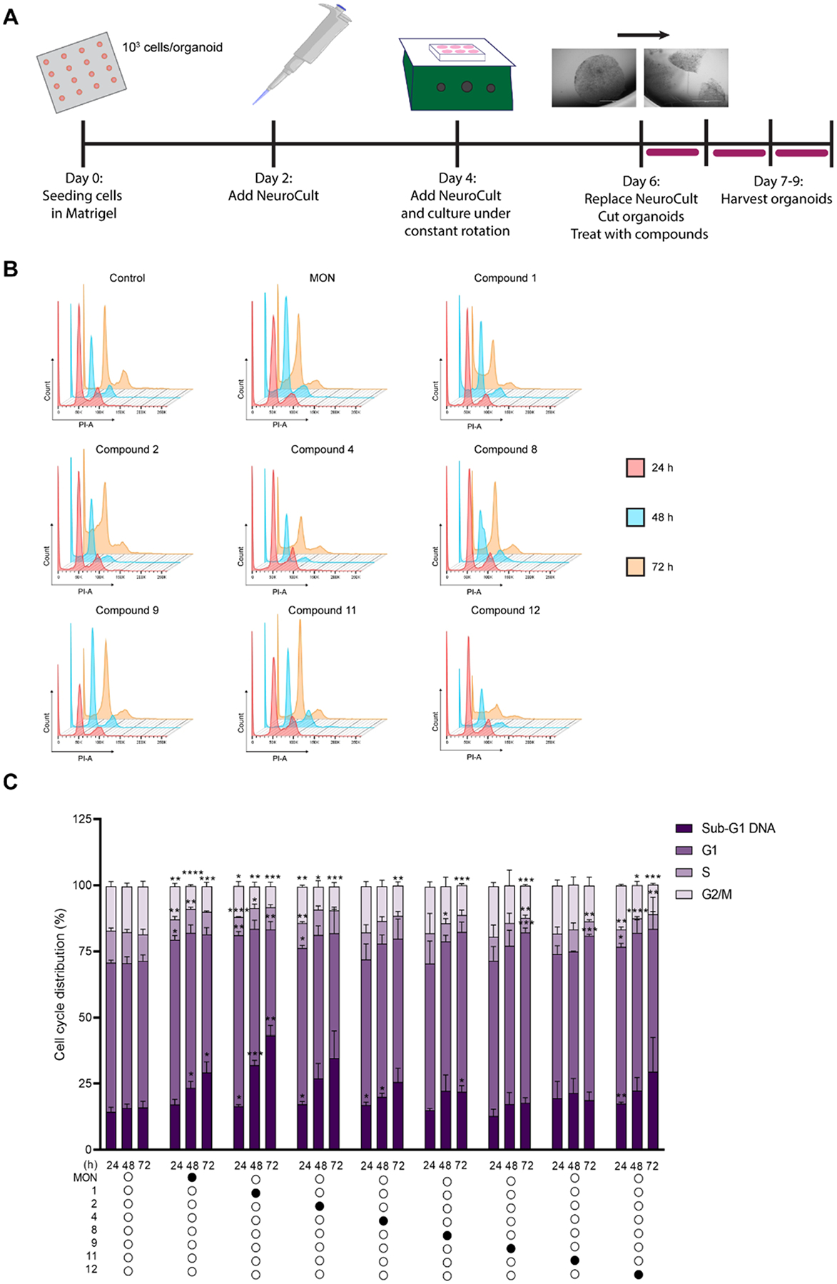
MON analogs induced DNA fragmentation in U-118MG organoids. U-118MG organoids were treated with 0.1% DMSO (control), parent MON or its select analogs at concentrations equal to 5 × IC 50 values for 24, 48 or 72 h and subsequently subjected to propidium iodide staining and flow cytometry. A. Schematic representation of U-118MG organoids generation and treatment; B. Representative cytograms; C. Distribution of cells in different phases of cell cycle or with Sub-G1 DNA determined by PI staining. Data represent mean ± SD (n = 6 for control, and n = 3 for compound treatment) *p ≤ 0.05, **p ≤ 0.01, *** p ≤ 0.001, ****p ≤ 0.0001.