
Fig. 7.
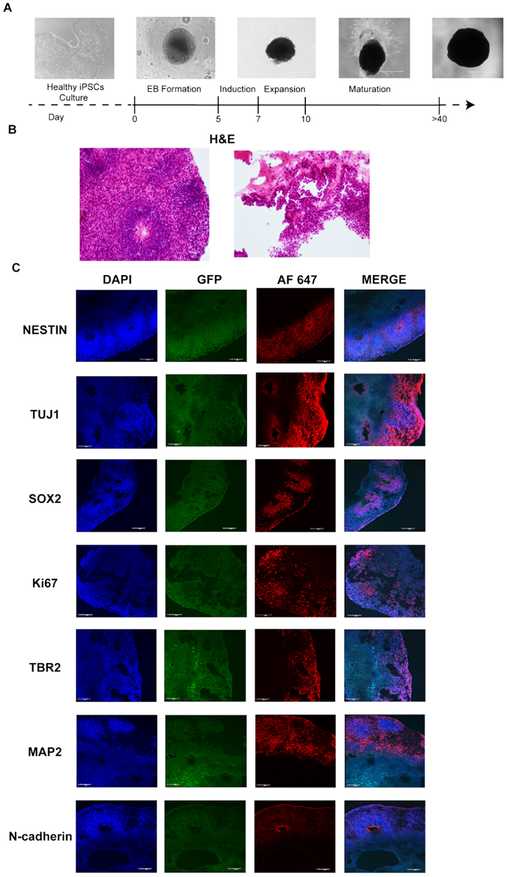
Cerebral organoids acquisition and validation. A. Schematic representation of cerebral organoids generation, scale bar 1000 μm; B. H&E staining of normal cerebral organoids at 51 days of development, 20X magnification; C. Immunofluorescence staining of DAPI, GFP and AF 647 – Alexa Fluor 647, a secondary antibody used for Nestin, Tuj1, Sox2, Ki67, TBR2, MAP2, N-cadherin, 20X magnification.