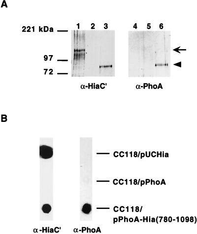
FIG. 4.
Immunoblot analysis of PhoA-Hia(780-1098) chimera. (A) Outer membrane proteins detected by Western blot with guinea pig antiserum against Hia residues 659 to 1098 in lanes 1 to 3 and with antiserum against PhoA in lanes 4 to 6. Lanes 1 and 4, E. coli CC118/pUC-Hia; lanes 2 and 5, E. coli CC118/pUC-PhoA; lanes 3 and 6, E. coli CC118/pPhoA-Hia(780-1098). Arrow indicates full-length Hia, and arrowhead indicates PhoA-Hia(780-1098). (B) Whole-cell immunoblots performed with either guinea pig antiserum against Hia residues 659 to 1098 (left lane) or antiserum against PhoA (right lane).

