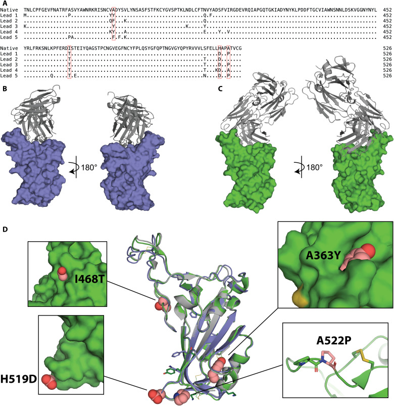
Fig. 3. Structural basis for immunogen stabilization.
(A) Sequence alignment of amino acid changes in lead immunogens relative to the native RBD sequence. Four recurring changes are highlighted in pink. (B) Crystal structure of lead 1 (slate) in complex with scFv C144 (gray). (C) Crystal structure of lead 3 (green) in complex with Fab P2B-2F6 (gray). (D) The crystal structures of lead immunogens 1 (slate) and 3 (green) are globally similar to WT RBD (gray; PDB:7BWJ) despite amino acid changes (recurring, spheres; unique, sticks). Insets illustrate key recurring substitutions (pink).

