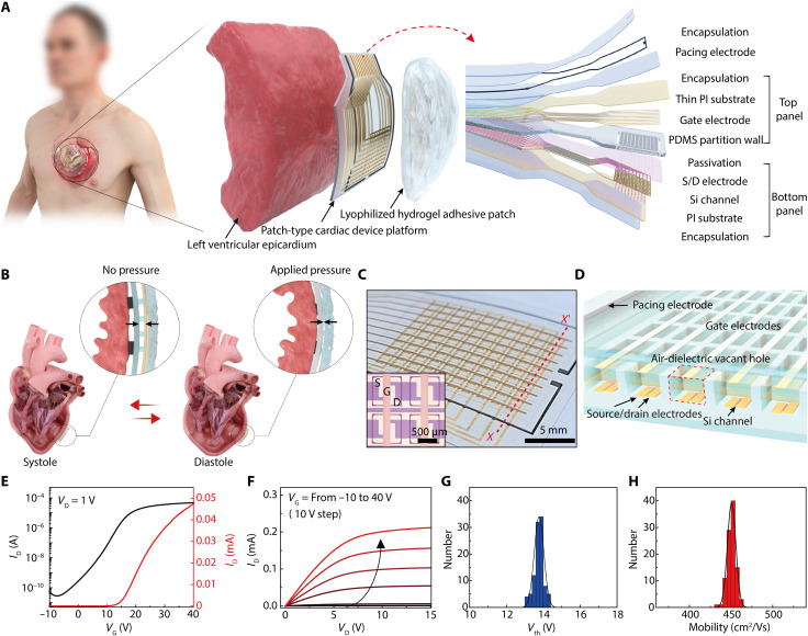
Fig. 1. Heart-attachable patch-type device platform.
(A) Schematic layouts of the device platform composed of an active-matrix pressure-sensitive transistor array and biocompatible pacing electrodes with encapsulation layers (right inset). This device platform was attached to the epicardium by covering a hydrogel adhesive patch for monitoring cardiac pressure and electrical treatment (left). (B) Schematic illustrations on the pressure sensing mechanism using this device platform during cardiac contraction (systole) and relaxation (diastole). (C) Photograph of the active-matrix pressure-sensitive transistor array and Pt black pacing electrodes (outer black lines) in the patch device platform. The 10 × 10 sensing nodes defined by elastomeric partitions are shown in the photograph. The inset shows an optical micrograph of four adjacent transistors that are isolated with elastomeric partitions. S, D, and G denote the source, drain, and gate electrodes, respectively. (D) Cross-sectional schematic illustration of the active-matrix air-dielectric transistor array [designated as X-X′ in (C)]. A red dashed box indicates the air-dielectric vacant hole that can be compressed when pressure is applied. (E and F) Representative transfer (VD = 1 V) and output (VG = −10 to 40 V) characteristics of the air-dielectric transistor in this epicardial device patch at ambient conditions. (G and H) Statistical distributions of the threshold voltage (Vth) and the field-effect n-channel mobility of 100 transistors in total, respectively.

