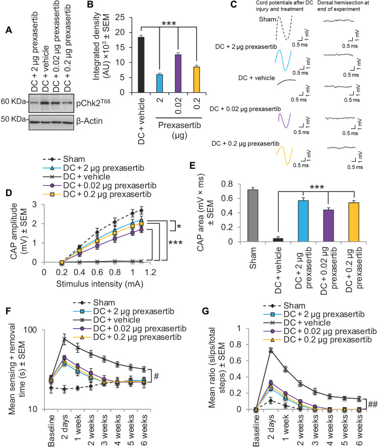
Fig. 7. Dose de-escalation study with prexasertib.
(A) Western blot and (B) densitometry to show that lower doses (0.2 and 0.02 μg) of prexasertib than that required to cause maximal suppression of pChk2 levels (i.e., 2 μg) were still able to significantly suppresses pChk2T68 levels after DC injury. (C) Spike 2 software–processed CAP traces from representative sham controls, DC + 2 μg prexasertib–, DC + vehicle–, DC + 0.02 μg prexasertib–, and DC + 0.2 μg–treated rats at 6 weeks after DC injury and treatment. Dorsal hemisection at the end of recording ablated all CAP traces. (D) Negative CAP amplitudes and (E) CAP area at different stimulation intensities were both significantly attenuated in DC + vehicle–treated rats but were dose-dependently restored in DC + prexasertib–treated rats [P = 0.00011, one-way ANOVA with Dunnett’s post hoc test (main effect)]. (F) Mean tape sensing/removal times and (G) mean error ratio to show the number of slips versus total steps are both restored to normal 3 weeks after treatment with 2 and 0.2 μg of prexasertib but were restored to normal 4 weeks after DC injury and treatment with 0.02 μg of prexasertib (#P = 0.00012, generalized linear-mixed models and ##P = 0.00014, linear-mixed models over the whole 6 weeks). A significant deficit remains in DC + vehicle–treated rats. n = 6 rats per treatment, three independent repeats (total, n = 18 rats per treatment).

