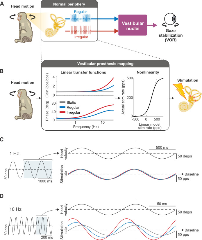
Fig 1. Biomimetic afferent response dynamics are implemented in the vestibular prosthesis mapping between head motion and stimulation rate.
(A) Schematic of the VOR pathway. Both types of vestibular afferents (regular and irregular) convey head movement information to the vestibular nuclei, which, in turn, stabilize the gaze via the VOR. (B) Schematic of how the prosthesis converts head movements into pulsatile stimulation. The mapping (black box) consists of the linear transfer functions, which mimic the response dynamics of regular (blue) and irregular (red) afferents or represent the conventional mapping with static gain and phase lead (gray), in cascade with the sigmoidal nonlinearity, which limits the firing rate to be above zero and below the maximum rate of 500 Hz. (C) Example of the stimulation rate, modulated around the baseline rate of 150 pps, in response to sinusoidal head movements at 1 Hz. At this frequency, the stimulation rates are similar for all mappings. Vertical line denotes the peak of head movement for phase comparison. (D) Example of the stimulation rate in response to sinusoidal head movements at 10 Hz. Both regular and irregular mappings display greater depth of modulation in firing and bigger phase lead than the static mapping, which remains in-phase and shows the same depth of modulation as in (C).

