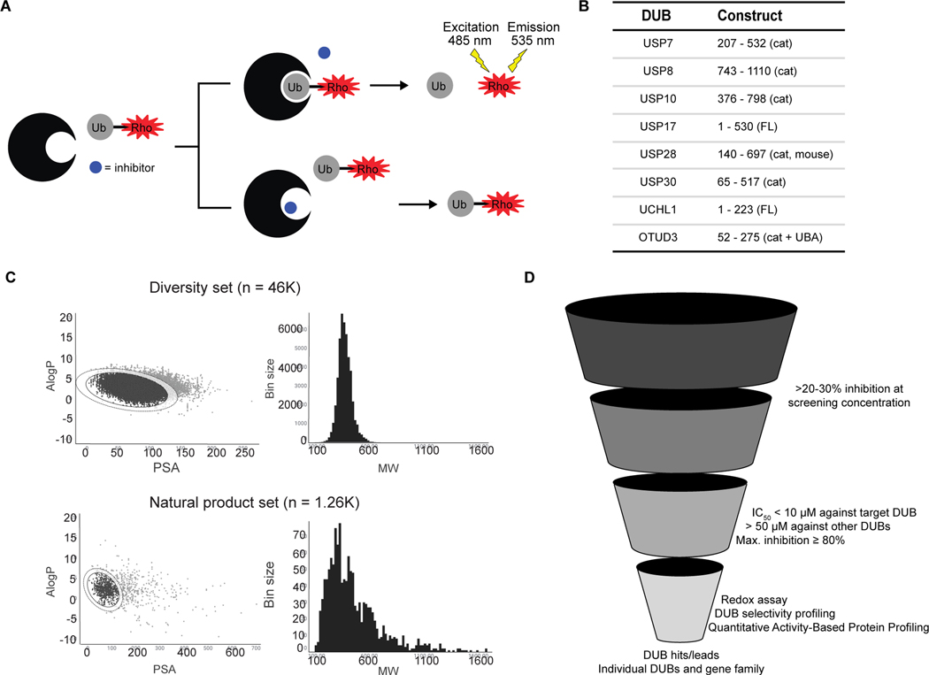
Figure 1. Overview of DUB HTS campaign.
A) Schematic of Ub-Rho screening assay. Uninhibited DUB cleaves rhodamine from ubiquitin (top) resulting in a fluorescent signal. In the presence of an inhibitor (bottom), the DUB cannot cleave the substrate, and fluorescence is unchanged.
B) DUBs included in the screen along with constructs used (cat denotes catalytic domain, FL denotes full length protein, cat + UBA denotes the catalytic domain plus UBA domain). All proteins are human isoforms except USP28, which is from mouse (mus musculus).
C) Summary of druglike properties partition coefficient (LogP) to polar surface area (PSA) and molecular weight (MW) among library members.
D) Diagram of screening cascade workflow.

