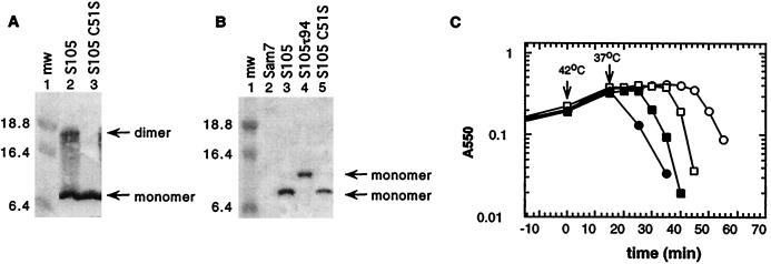
FIG. 2.
S monomer and dimer formation in the cytoplasmic membrane. (A) Membrane protein samples were prepared from MC4100(λKnΔSR) + pS105 (lane 2) or MC4100(λKnΔSR) + pS105C51S (lane 3) and analyzed by Western blotting. (B) Membrane protein samples of MC4100(λCmΔSR) bearing the plasmids pKB1 (Sam7) (lane 2), pS105 (lane 3), pS105τ94 (lane 4), or pS105C51S (lane 5) were prepared in the presence of NEM in the membrane extraction buffer and analyzed by Western blotting. Molecular weights (mw) of the prestained molecular standards (in thousands) (lane 1) are indicated to the left. S monomer and dimer bands are indicated by arrows. (C) MC4100(λKnΔSR) cells carrying the plasmids pKB110 (S+) (○), pS105 (□), pSC51S (●), or pS105C51S (■) were induced and monitored for turbidity.

