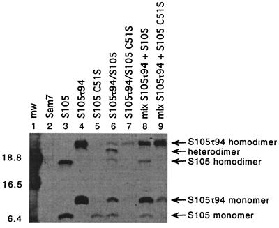
FIG. 3.
S dimer formation in the bacterial membrane. Oxidation and sample preparation for Western blot analysis was performed as described in Materials and Methods. Samples were prepared from the indicated strains: lane 1, molecular weight standards; lane 2, MC4100(λCmΔSR) + pKB1 (Sam7); lane 3, MC4100(λCmΔSR) + pS105; lane 4, MC4100(λCmΔSR) + pS105τ94; lane 5, MC4100(λCmΔSR) + pS105C51S; lane 6, MC4100(λCmS105τ94) + pS105; lane 7, MC4100(λCmS105τ94) + pS105C51S; lane 8, mixed cultures MC4100(λCmΔSR) + pS105τ94 and MC4100(λCmΔSR) + pS105; lane 9: mixed cultures MC4100(λCmΔSR) + pS105τ94 and MC4100(λCmΔSR) + pS105C51S. Molecular weights of the prestained molecular standards (mw) (in thousands) are indicated to the left of the panel. S monomer and dimer bands are indicated with arrows.

