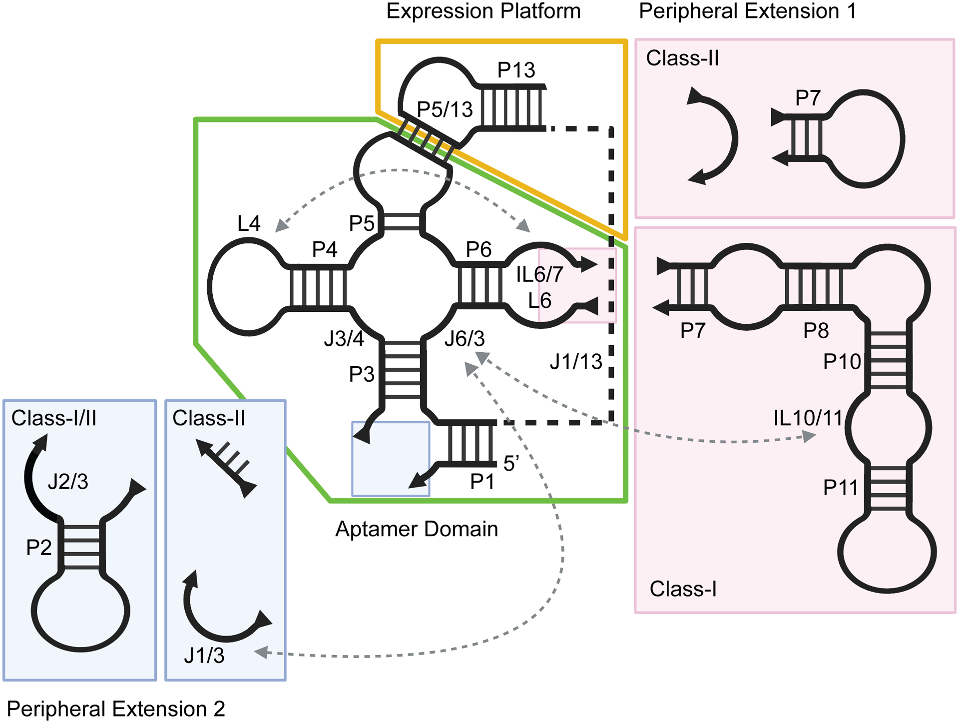
Figure 2.

Secondary structure schematic of cobalamin riboswitches, delineating structural differences between class-I and class-II. The conserved aptamer domain is boxed in green and the expression platform in yellow. Peripheral extensions which vary between classes are colored in pink (subdomain 1) and blue (subdomain 2). Long-range tertiary contacts are indicated by dashed arrows. Created with Biorender.com.
