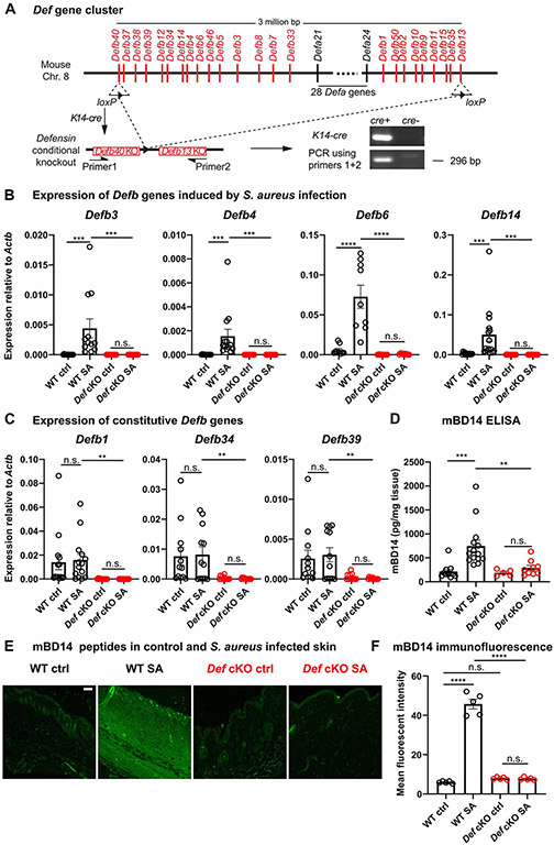
Figure 2. Generation of Defensin cluster conditional knockout mice.
(A) Schematic illustration of the Def gene cluster on mouse Chromosome 8. Two loxP sites were inserted into exons of Defb40 and Defb13 on the two ends of the gene cluster. K14-cre-mediated recombination successfully removed the entire 3 million base pair genomic region from keratinocytes. Genotyping PCR using primers flanking the cluster detected a band corresponding to partial sequence in Defb40, one recombined loxP site, and partial sequence in Defb13.
(B-C) mRNA quantification of infection-induced Defb3 (n=12), Defb4 (n=13), Defb6 (n=9) and Defb14 (n=15) (B) and constitutively expressed Defb1 (n=14), Defb34 (n=12) and Defb39 (n=12) (C) in WT (black) and Def cKO (red) control (Ctrl) skin and 24 hours post S. aureus infection (SA) by qPCR. The amounts of mRNA for each gene were normalized to housekeeping gene Actb.
(D) Quantification of mBD14 peptide in control and S. aureus infected skin of WT and Def cKO animals by ELISA. n=5-14
(E) Representative images of mBD14 immunofluorescence of WT control skin, WT skin infected with S. aureus, Def cKO control skin and Def cKO skin infected with S. aureus. mBD14 was low in uninfected WT skin but was robustly induced 24 hours after infection by S. aureus. In Def cKO animals, infection failed to induce mBD14 production. Scale bar=100μm
(F) Quantification of mean fluorescent intensity of anti-mBD14 immunofluorescence across the thickness of the skin tissue as shown in (E). n=5
Results are presented as mean ± SEM from at least three independent experiments. **p < 0.01, ***p < 0.001, ****p < 0.0001 n.s. not significant by one-way ANOVA.
See also Figure S2.

