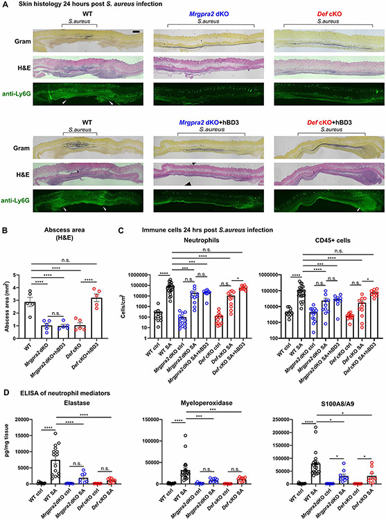
Figure 5. Mrgpra2 dKO and Def cKO animals showed severe defects in neutrophil abscess formation following S. aureus infection.
(A) Top row: Histology staining of WT, Def cKO, and Mrgpra2 dKO mouse skin 24 hours post S. aureus infection using Gram stain (top), H&E (middle), and immunofluorescent staining using a monoclonal antibody against Ly6G (bottom) to show the locations of neutrophils. Black brackets indicate the bacteria band. White arrowheads point to neutrophil abscesses. Scale bar=1mm
Bottom row: hBD3 injection rescued abscess formation in Def cKO but not Mrgpra2 dKO mutant animals.
(B) Quantification of abscess areas based on H&E staining shown in (A). n=5
(C) Flow cytometry analyses of total CD45+ cells and neutrophils in the skin 24 hours post S. aureus infection. Both Def cKO and Mrgpra2 dKO animals showed reduced recruitment of neutrophils. hBD3 injection rescued neutrophil recruitment of Def but not Mrgpra2 mutants. n=9-23
(D) ELISA quantifications of elastase, myeloperoxidase (MPO), and calprotectin (S100A8/A9 dimer) in the skin of WT, Def cKO and Mrgpra2 dKO mice 24 hours post S. aureus infection. n=6-21
Results are presented as mean ± SEM from at least three independent experiments. *p < 0.05, ***p < 0.001, ****p < 0.0001, n.s. not significant by one-way ANOVA.
See also Figure S5.

