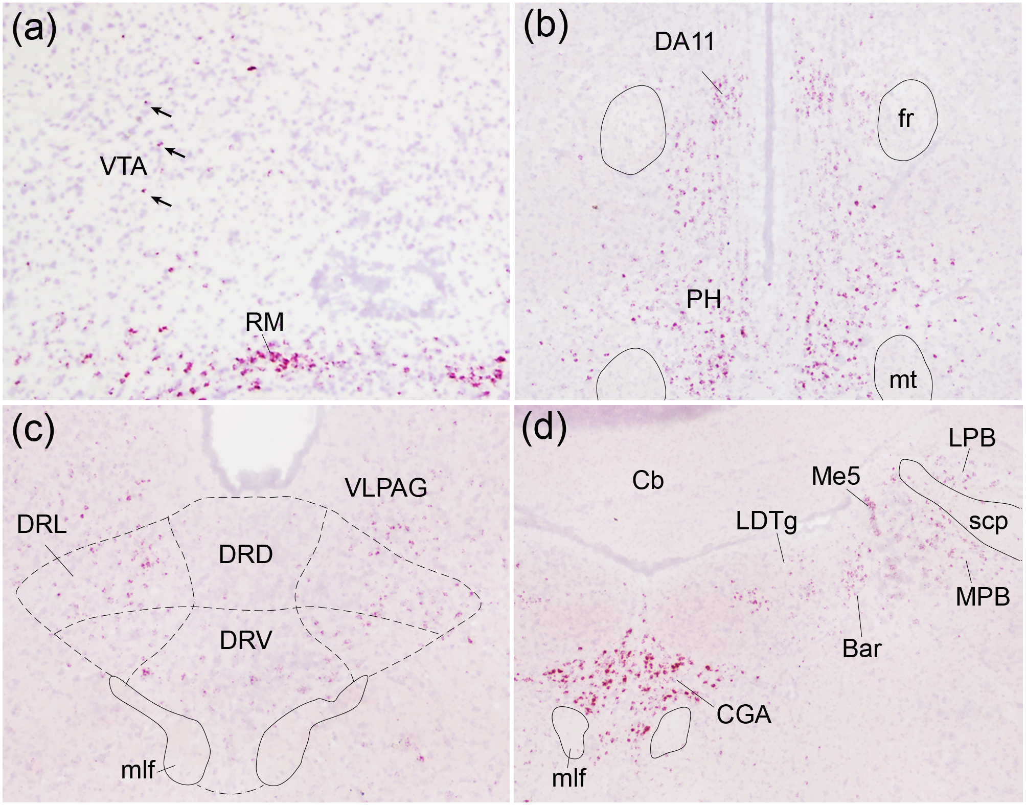
Figure 5. Oxtr expression in other regions of interest.

Oxtr mRNA (red) was labeled with in situ hybridization against a Hematoxylin counterstain (purple). The ventral tegmental area (VTA; a) and dorsal raphe nucleus (DR; c) displayed spare Oxtr labeling while the retromammillary nucleus (RM; a), posterior hypothalamic nucleus (PH; b), and central gray, alpha part (CGA; d) displayed robust labeling. Arrows indicate Oxtr-positive cells within VTA; DA11, DA11 dopamine cells; fr, fasciculus retroflexus; mt, mammillothalamic tract; DRD, dorsal raphe nucleus, dorsal part; DRL, dorsal raphe nucleus, lateral part; DRV, dorsal raphe nucleus, ventral part; VLPAG, ventrolateral periaqueductal gray; mlf, medial longitudinal fasciculus; CG, central gray, LDTg, laterodorsal tegmental nucleus; LPB, lateral parabrachial nucleus; MPB, medial parabrachial nucleus; Me5, mesencephalic trigeminal nucleus; Bar, Barrington’s nucleus; Cb, cerebellum; scp, superior cerebellar peduncle.
