
Figure 6. Oxtr mRNA and OXTR protein mismatch in hippocampus.
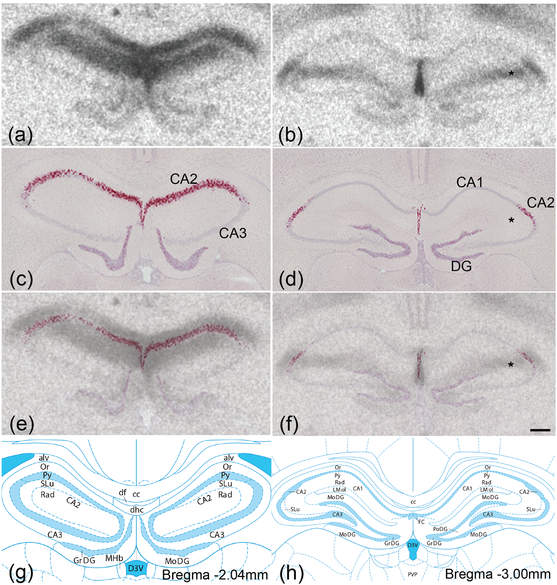
OXTR protein (black; a,b) was labeled with autoradiography and, in adjacent sections, Oxtr mRNA (red; c,d) was labeled with in situ hybridization against a Hematoxylin counterstain (purple). Images from adjacent autoradiography and in situ hybridization sections were merged (e,f) to compare protein and mRNA localization. Oxtr mRNA was restricted to stratum pyramidale, the principal cell layer containing the somas of pyramidal cells, while robust OXTR protein binding was present superficial and basal to stratum pyramidale, most likely in the stratum radiatum, stratum lacunosum-moleculare, and stratum oriens. Illustrations from the Rat brain Atlas indicate that Oxtr mRNA was enriched in the CA2 in the Ammon’s horn (g,h). DG, dentate gyrus; * denotes OXTR protein in the absence of Oxtr mRNA. Scale bar =300μm shown in (f).