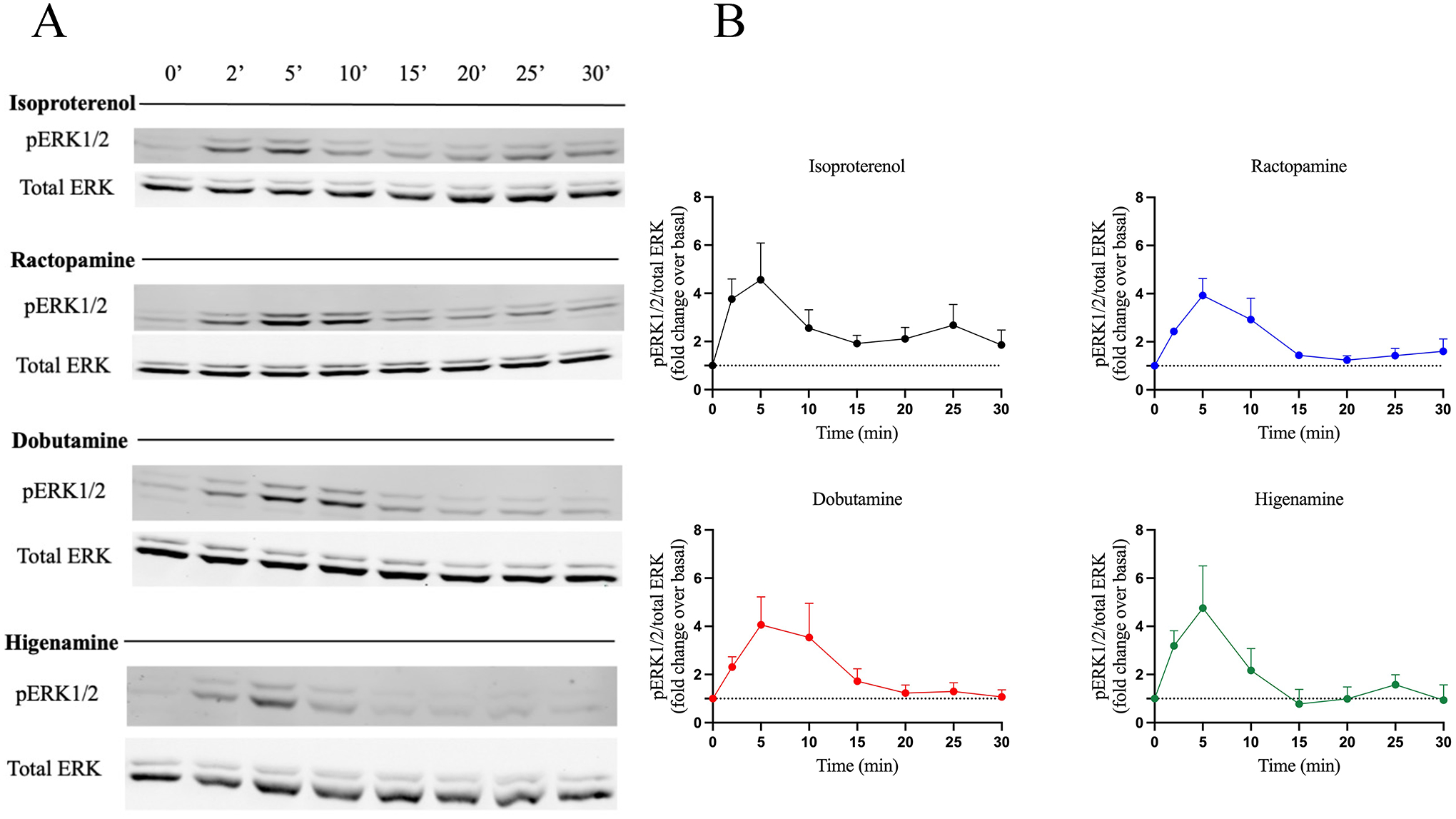
Figure 4 – Activity of β-agonists on ERK phosphorylation.

ERK phosphorylation was assessed by western blotting and results are shown as (A) a representative western blot from three independent experiments and (B) normalized data after quantification using ImageJ. HEK293 cells endogenously expressing the β2AR were stimulated with 10 μM ISO, RAC, DOB or HIG for 0, 2, 5, 10, 15, 20, 25 and 30 min. Cells were lysed and equal amounts of protein were analyzed by western blot to detect ERK1/2 phosphorylation. Phopho-ERK1/2 (pERK1/2) bands were quantified and normalized to total ERK1/2 (pERK/total ERK) and are represented as fold change over basal. Data are mean ± SEM, n = 3.
