
Figure 1.
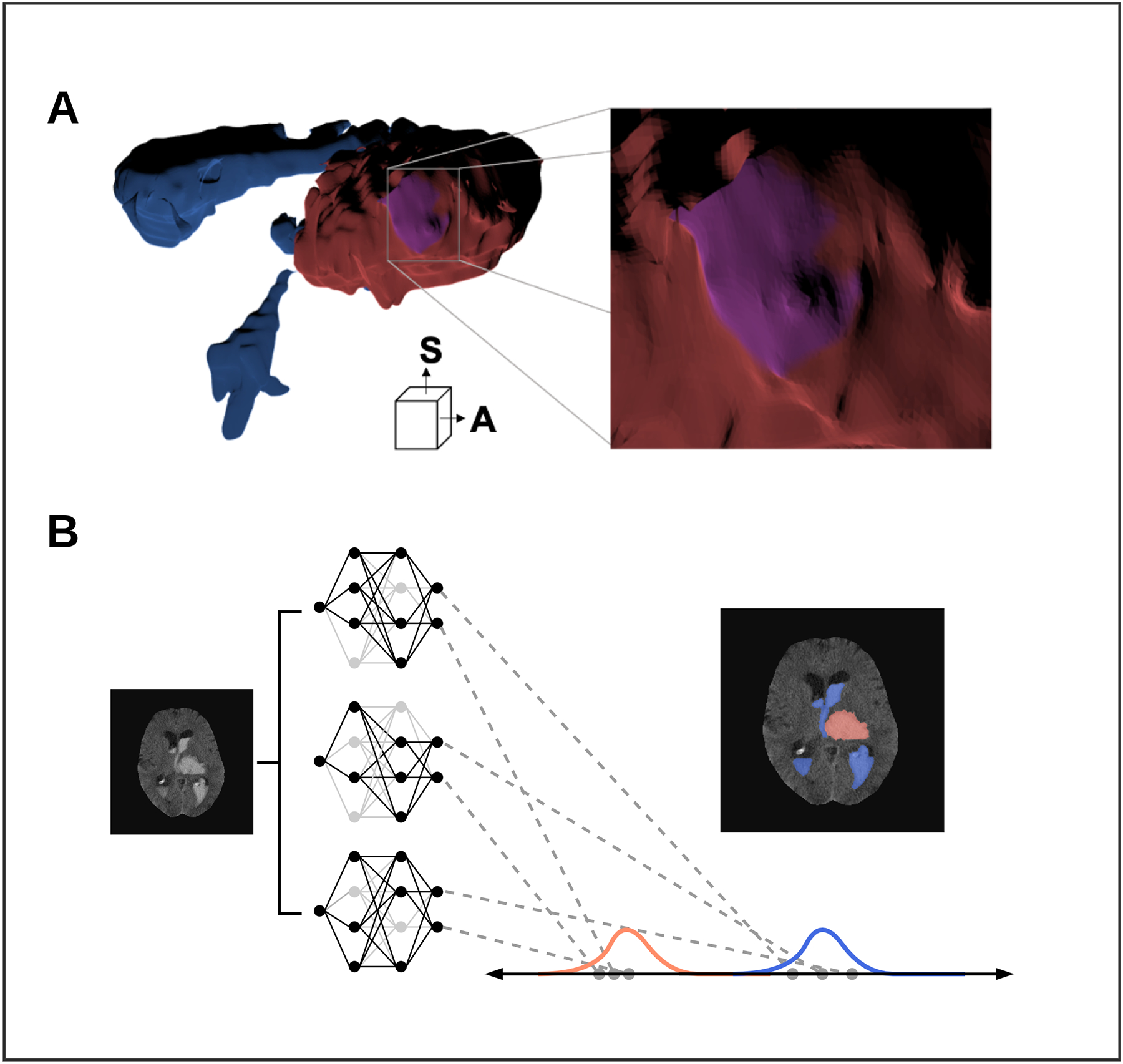
Advanced imaging techniques including, ventricular intersection area (VIA) mesh construction and Bayesian deep learning uncertainty approximation.
A) 3D inner-surface renderings of ICH (Red), IVH (Blue) and the VIA (Purple) generated by a single rater. Anterior (A) and superior (S) directions are displayed Right: zoomed portion of the VIA along the inner ICH surface. B) Bayesian approximation in a deep neural network by Monte-Carlo dropout. Networks are loaded with different nodes stochastically ‘dropped out’ (light-grey) to create a group of related networks. Each segmentation is a distribution of ICH (red) and IVH (blue) segmentations.