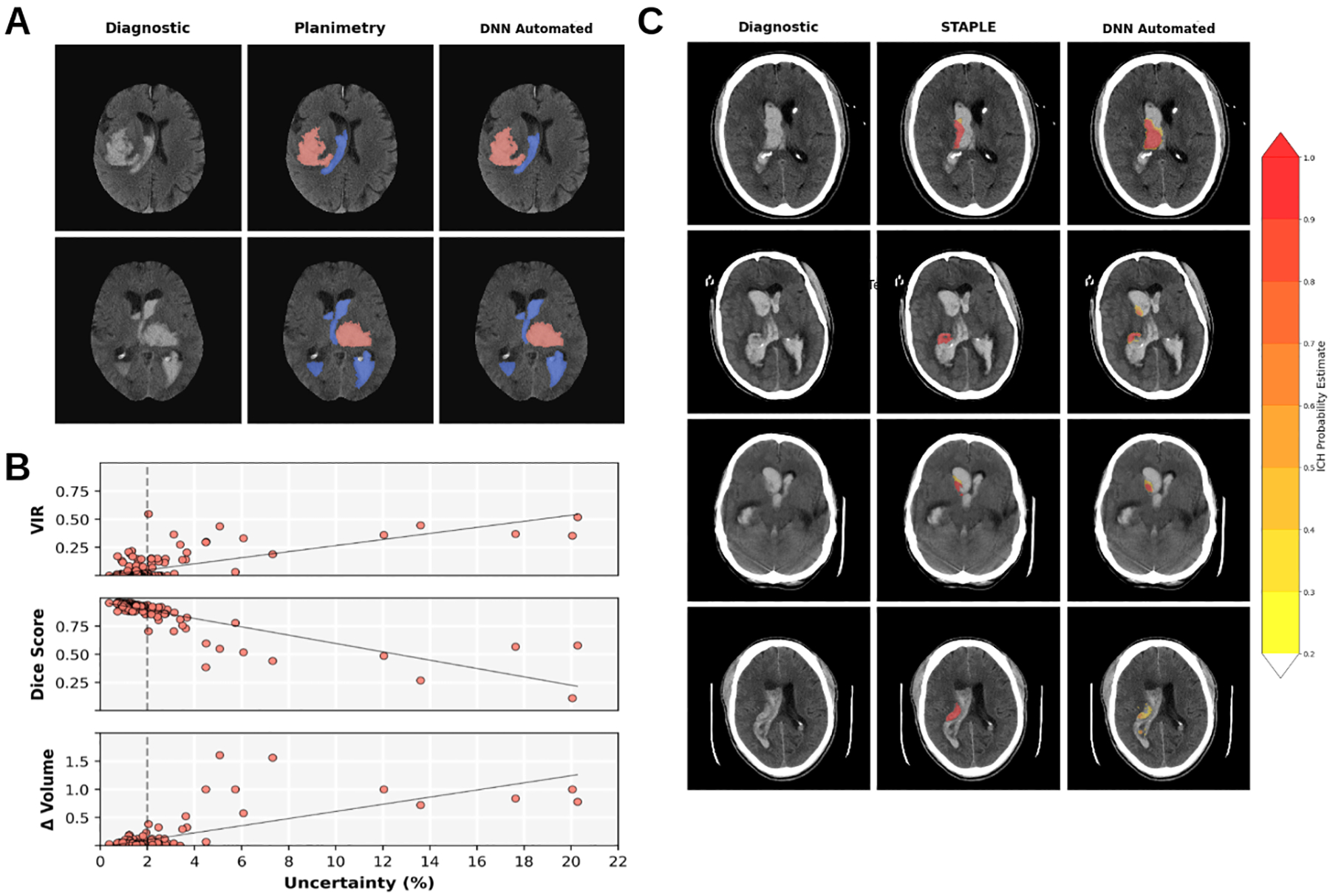
Figure 4.

Bayesian Deep Neural Network (DNN) segmentation images and uncertainty vs segmentation quality analysis. A) Median Dice score DNN automated segmentation images from MISTIE II (first row) and CLEAR IVH (second row) with ICH (red) and IVH (blue). B) Segmentation quality metrics vs Uncertainty (%) for majority voting ICH segmentations by from MISTIE II and CLEAR IVH. Top: Ventricular intersection ratio (VIR) vs Uncertainty (r= 0.699). Middle: Dice Scores vs Uncertainty (r=− 0.849). Bottom: The relative volume difference (Δ Volume) vs Uncertainty. For values beyond 2% (dashed lines) we see deviations from the linear relationships. C) Simultaneous truth and performance level estimation (STAPLE) and DNN Automated probability estimates of four patients with VIR > 0.25. On the left is the diagnostic scan, the center is overlayed with the planimetry STAPLE consensus estimate and right the DNN probabilistic output. Far right is a probability estimate colormap.
