Figure 3.

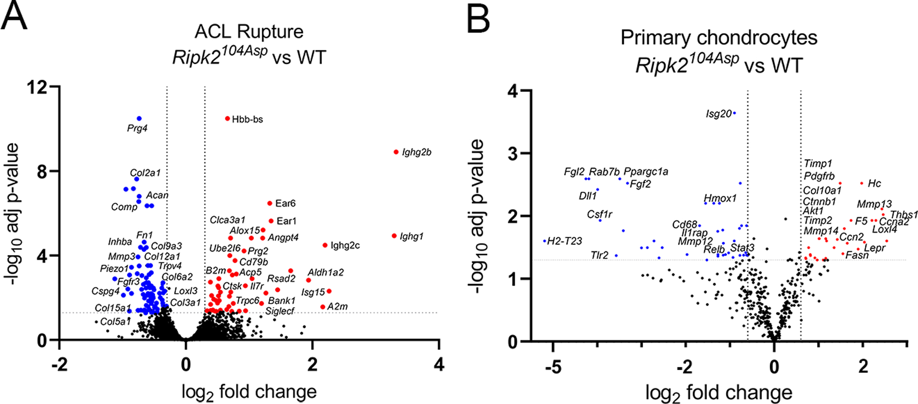
Ripk2104Asp enhances expression of OA-associated markers in the surgically injured joint as well as primary chondrocytes. (A) Comparative analysis of RNA-seq performed on whole joints isolated from WT or Ripk2104Asp mice 10 days post ACL rupture. The volcano plot indicates genes significantly upregulated (red) or downregulated (blue) in Ripk2104Asp as compared to WT joints. (B) The nCounter Fibrosis panel was used to measure gene expression in AC cultured from WT or Ripk2104Asp mice. Volcano plot indicates genes significantly upregulated (red) or downregulated (blue) in Ripk2104Asp primary chondrocytes compared to WT primary chondrocytes.
