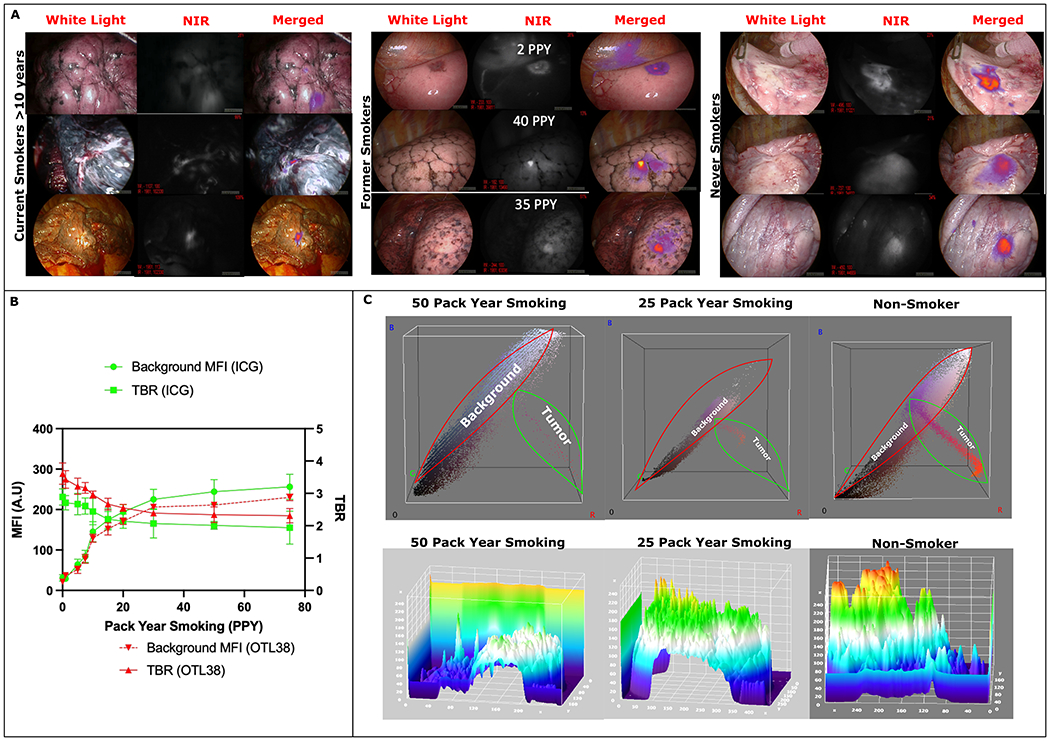
Figure 2:

(A): Left- Representative white light, NIR, and merged images from patients who were current smokers with >10 PPY at the time of surgery. As seen background lung parenchyma is significantly darker from smoke related inflammation leading to decreased contrast between tumor and background. Middle: representative images from past smokers showing background parenchymal inflammation are correlated with PPY. Right Representative images from never smokers showing minimal normal parenchyma inflammation and excellent contrast between normal lung and the tumor.
(B): Trend of background parenchymal intensity in relation to patient pack year smoking showing increased NIR fluorescence leading to lower TBR measurements in patients with higher smoking exposure.
(C): Top Row: 3D RGB analysis shows increased background color uptake than tumor fluorescence in patients with smoking history. Fused NIR images of tumors have increased Red color emission which is minimal in the 50 PPY group whereas Red color emission is significantly elevated in the non-smoker group highlighting increased tumor fluorescence and lower background detection.
Bottom Row: 3D Topographical heat map showing decreased tumor intensity compared to background lung parenchymal fluorescence (i.e increased background fluorescence dampening tumor fluorescence intensity as detected by the device) in the smokers compared to non-smokers.
