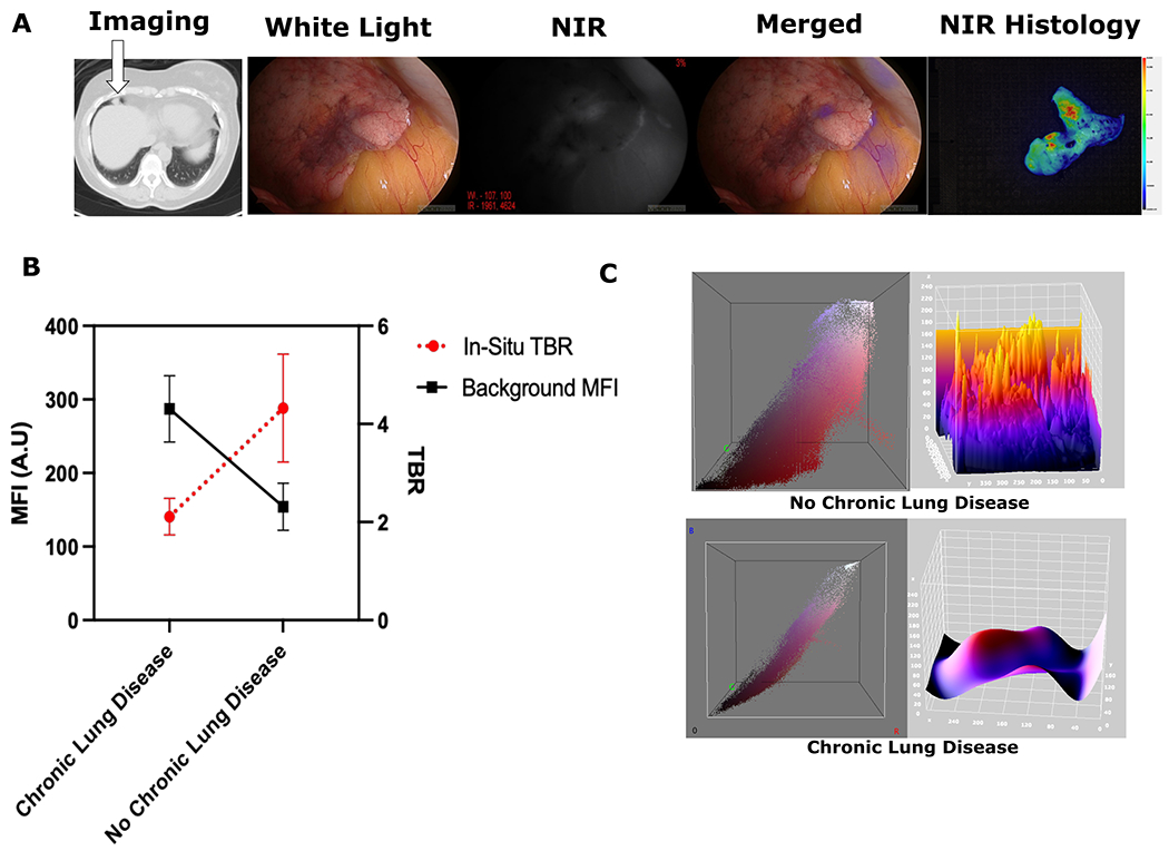
Figure 3:

(A): Sample images from IMI guided resection of lung lesion from a patient with 20 year history of chronic bronchitis. The lesion was hard to localize intra-operatively due to poor delineation from surrounding tissues, but IMI histology shows NIR tracer localization in the nodule. (B): Comparison of the background MFIs for Chronic Lung Disease patients vs those No Chronic Lung Disease shows increased background signal in those with chronic lung conditions (p<0.05). Similar statistically significant observation was also noted for TBR measurements between the groups (p<0.05) (C): Representative 3D analysis of an IMI guided resection showing increased fluorescence intensity in lung parenchyma in RGB and 3-D topographic analysis for chronic lung disease patients vs non chronic lung disease.
