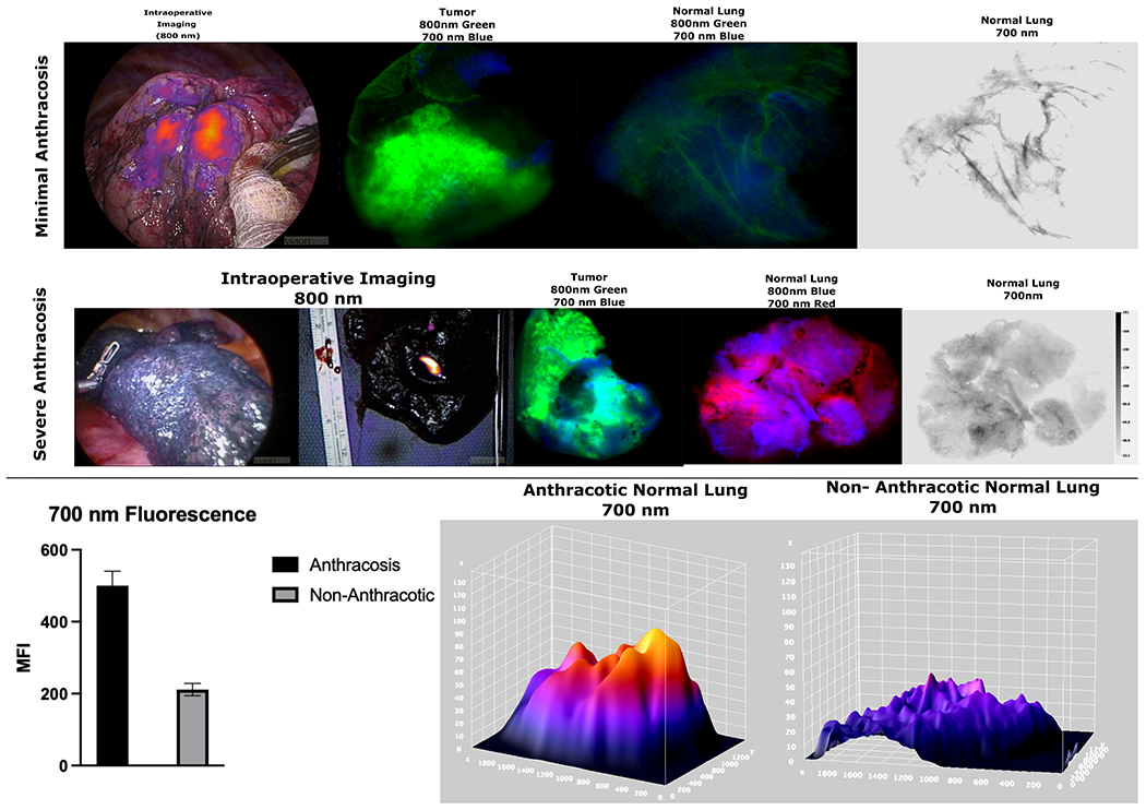
Figure 6:

Top Row: Specimen analysis of a minimally anthracotic lung during IMI guided cancer resection shows excellent tumor localization with ICG. Specimen analysis shows high tumor fluorescence in 800 nm wavelength with minimal background fluorescence and minimal fluorescence when analyzing the 700 nm wavelength emission.
Middle Row: Patient with severe anthracosis who underwent OTL38 guided lung cancer resection with specimen analysis showing very elevated fluorescence of normal tissue at 700 nm range. It should be noted that during in-vivo imaging during IMI, there was no fluorescence observed (left image) and lesion only fluoresced after the nodule was bisected demonstrating the limitations on tumor nodule fluorescence emission the anthracotic parenchyma presents
Bottom Row: Lungs with severe anthracosis due to light absorbing carbons showed statistically elevated emission at 700 nm (p<0.05) with 3-D analysis showing elevated background MFI at those wavelengths as well.
