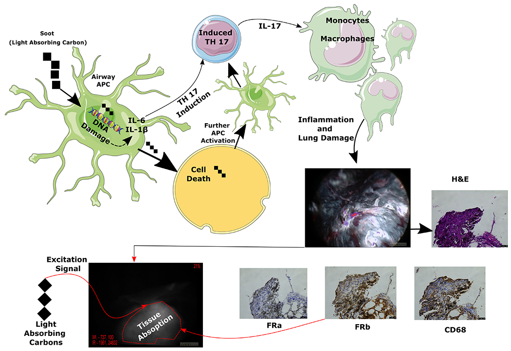
Figure 7:

Pictorial representation of two mechanisms involved in anthracosis related background lung fluorescence. It is thought that soot (light absorbing carbons) gets taken up by antigen presenting cells (APC) which causes DNA breaks leading to cell death and induction of inflammatory cytokines which activated the adaptive immune system leading to inflammatory cascade. One of the cells that is recruited during this inflammatory response are the monocytes which become macrophages (see CD68 stain) and stain for folate receptor beta which is thought to bind folate targeted NIR tracers albeit at a lower degree. Additionally, soot because of its innate properties leads to black pigmentation and absorbs light/energy at a higher rate. Combination of these factors is thought to contribute to increased fluorescence of anthrocotic lung tissue during IMI.
