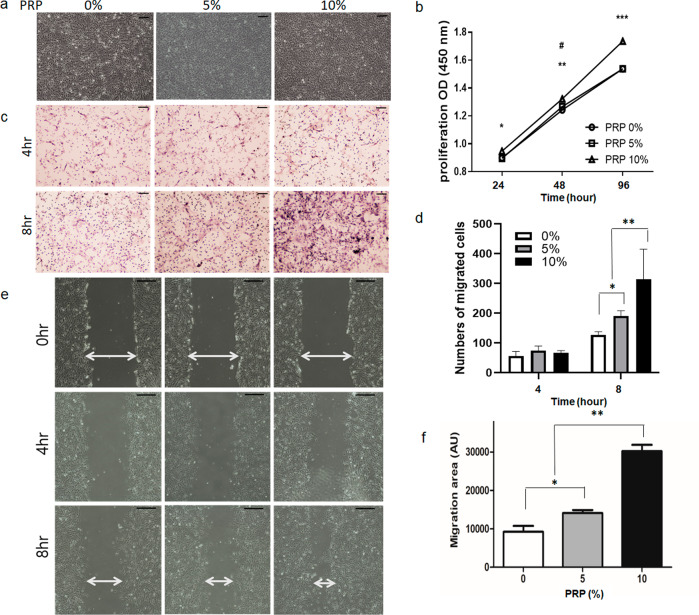
Fig. 4. Analysis of the proliferation and migration of human SCs.
a human SCs exhibited a spindle-shaped and bipolar or occasionally multipolar cell morphology. As the concentration of PRP was increased, the proliferation of human SCs increased. (scale bar, 100 μm). b The number of human SCs were increased significantly and dose-dependently by PRP. After treatment for 96 hours, 10% PRP increased SC cell count significantly more than in non-treated controls. (#, compared between 0% and 5%, #P < 0.05; *compared between 0% and 10%, *P < 0.05, **P < 0.005, **P < 0.0001). c Representative light photomicrographs of migrated human SCs induced by 0%, 5%, and 10% PRP after incubation for 4 or 8 h by the transwell assay (scale bar, 100 μm). d Human SCs treated with 5% PRP for 8 h had significantly greater proliferation rates than non-treated controls (*P < 0.05) The number of migrated cells in 10% PRP is higher than in both non-treated cells and 5% PRP treated cells. (**P < 0.0001, respectively). e PRP significantly and dose-dependently increased SCs migration rate as compared with non-treated SCs. f The migration rate of cells treated with 10% PRP was 4.25-fold greater than that of non-treated controls (P < 0.05). SCs Schwann cells, PRP platelet-rich plasma.

