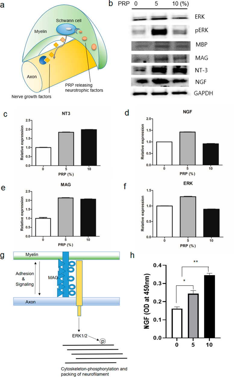
Fig. 5. In vitro neurotrophic factor secretion behavior after PRP treatment on human SCs.
a Schematic illustration of nerve. An axon surrounded by a myelin sheath and Schwann cells, which are located in myelin and release neurotrophic factors. b Western blot analysis of neural regeneration-related proteins in human SCs which were treated with different concentrations of PRP. All blots derive from the same experiment and were processed in parallel. c, d Quantification of neurotrophic factors including NT3 and NGF. As the concentration of PRP increased the expressions of neurotrophic factors increased more so than that observed in non-treated controls. e, f MAG expression was increased dose-dependently by PRP. ERK (a MAG-related axon cytoskeletal protein) expression was also increased by PRP. g Schematic illustration of connection between axon and myelin. h The expression of NGF in human SCs by ELISA; human SCs were observed to express NGF in a PRP dose-dependent manner. SC Schwann cell, PRP platelet-rich plasma, NGF nerve growth factor, NT-3 neurotrophin-3, MAG myelin-associated glycoprotein, MBP myelin basic protein, ERK extracellular signal-regulated kinase protein.

