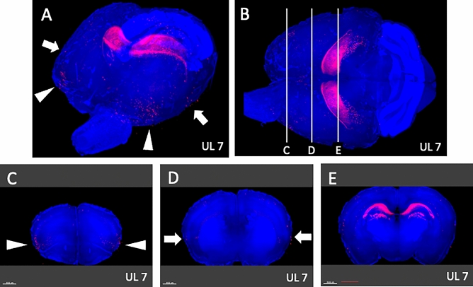
Figure 10.
Survey on arbitrary cross sections on UL7. (A) Panoramic view of whole-brain imaging on day 7 after UL. Neural activity can be observed in the anterior-temporal (arrowheads) and temporal (arrows) regions. (B) Images from the top of the brain. The vertical lines indicate the cross-sectional positions in panels (C–E). (C) The activated neurons in the anterior temporal region (arrowheads, panel A) are located in the agranular insular cortex (AIC) (arrowheads). (D) Activated neurons in the temporal region (arrows, panel A) were located in the primary somatosensory cortex (S1). (E) Activated neurons observed in the caudate putamen on day 2 were decreased on day 7 after UL. Activated neurons in the caudate putamen were not observed.

