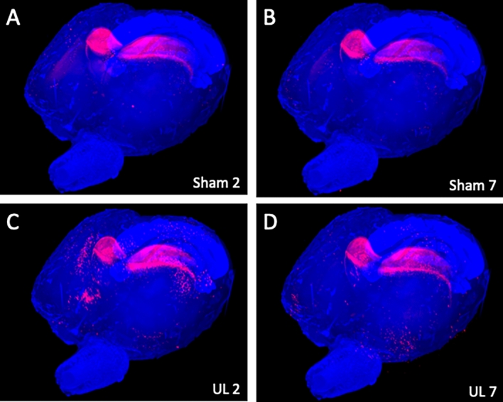
Figure 2.
Representative whole-brain images of each group. Panoramic view of whole-brain imaging using CUBIC analysis. Red fluorescence indicates activated neurons. There were many non-specific activated neurons in the hippocampus in all images. (A) Images obtained 2 days after sham surgery. (B) Images obtained 7 days after sham surgery. (C) Image obtained 2 days after unilateral (left) labyrinthectomy (UL). (D) Image obtained 7 days after UL.

