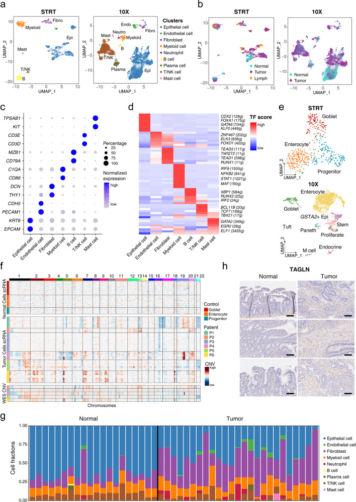
Fig. 1. Expression landscape of SBA and cell composition changes.
a UMAP plot exhibiting the identified clusters of the STRT and 10× datasets. b UMAP plot exhibiting the cell tissue sources of the STRT and 10× datasets. c Dot plots presenting the normalized expression level of corresponding markers of each cell cluster in the STRT dataset. d Heatmap showing the average normalized GRN expression scores. e UMAP plot exhibiting the identified clusters of normal epithelial cells in the STRT and 10× datasets. f The upper panel exhibiting large-scale CNVs of single cells inferred based on normal epithelial cells in the STRT dataset. The middle panel exhibiting large-scale CNVs of single cells inferred based on epithelial cells from tumor tissues in the STRT dataset. The lower panel exhibiting the CNV called from the WES data of P3–P6. g Histogram showing the cell composition percentage of each sample inferred by the deconvolution analysis. h Representative IHC staining of TAGLN in adjacent normal and primary tumor tissues (original magnification 100×). The rows of the paired normal and tumor samples are from the same patients, and three individual patients are listed. Scale bar, 100 μm.

