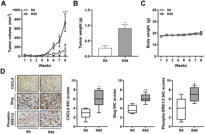
Figure 5.
Enhanced breast tumor growth and protein expression of CXCL5, Slug, and ERK phosphorylation in mice via xenograft of breast cancer cells after co-culture with resistin-stimulated ADSCs. The isolated ADSCs were treated with resistin at 0 and 50 ng/ml (denoted as R0 and R50, respectively) for 48 h, followed by co-culture with MDA-MB-231 cells in the transwell model for another 72 h. After the co-culture, MDA-MB-231 cells were collected and injected into the fourth mammary fat pads of female NOD/SCID mice for the following analyses. (A) The tumor volume, calculated by (width2 × length)/2, was measured weekly after palpable tumor mass was formed. (B) Upon sacrifice of the mice on week eight, the weight of individual tumor mass was measured. (C) The body weight of the mice was measured weekly. (D) The tumor mass was collected after sacrifice of the mice on week eight, and analyzed for CXCL5, Slug, and phospho-ERK1/2 protein expression by immunohistochemistry (IHC). The quantitative IHC scores were manually evaluated and calculated by multiplying the categorized percentage of stained cells (0, 0–24%; 1, 25‑49%; 2, 50‑74%; 3, 75‑100%) by the categorized intensity of staining (0, negative; 1, weak; 2, moderate; 3, strong). Data were obtained from three to six mice in each group and presented as mean ± SEM or box plots. Statistical difference was determined by t-test comparing R50 group versus R0 group as control. *p < 0.05; **p < 0.01; ***p < 0.001.

