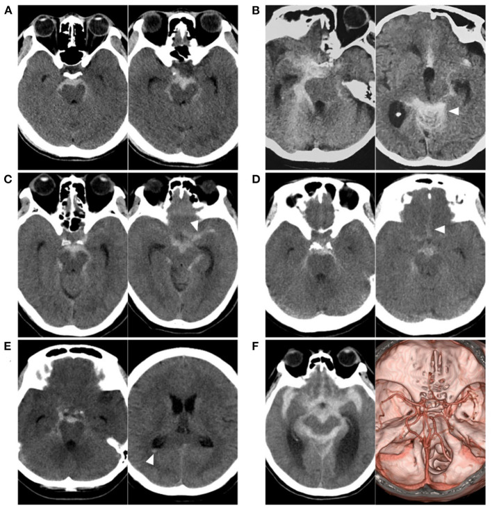
Figure 1.
Various SAH with negative angiography. (A) CT showing a PNSAH only with focal pre-pontine hemorrhage. (B) CT showing a PNSAH with involvement of quadrigeminal cistern (arrowhead). (C) CT showing a PNSAH extended to the basal part of the Sylvian fissure (arrowhead). (D) CT showing a PNSAH extended to the posterior part of the interhemispheric fissure (arrowhead). (E) CT showing a PNSAH with intraventricular sedimented blood (arrowhead). (F) Left: CT showing a diffuse non-PNSAH; Right: the intracranial aneurysm was not found on CTA; the drainage pattern of the deep vein around vein of Galen was normal. CT, computed tomography; CTA, computed tomography angiography; PNSAH, perimesencephalic non-aneurysmal subarachnoid hemorrhage; SAH, subarachnoid hemorrhage.

