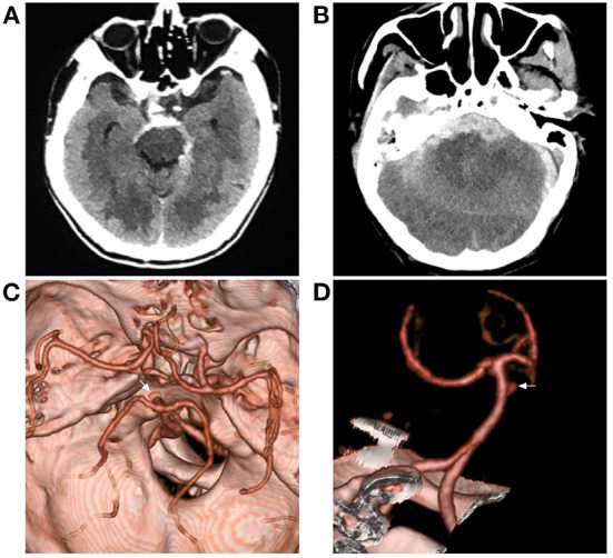
Figure 2.

Aneurysmal SAH with perimesencephalic bleeding pattern. (A) Immediate CT of the onset showing SAH with typical perimesencephalic bleeding pattern. (B) Repeated CT 6 h after the onset showing a second SAH with the thick hemorrhage in front of the brainstem. (C,D) CTA showing an aneurysm of the basilar artery (arrows). CT, computed tomography; CTA, computed tomography angiography; SAH, subarachnoid hemorrhage.
