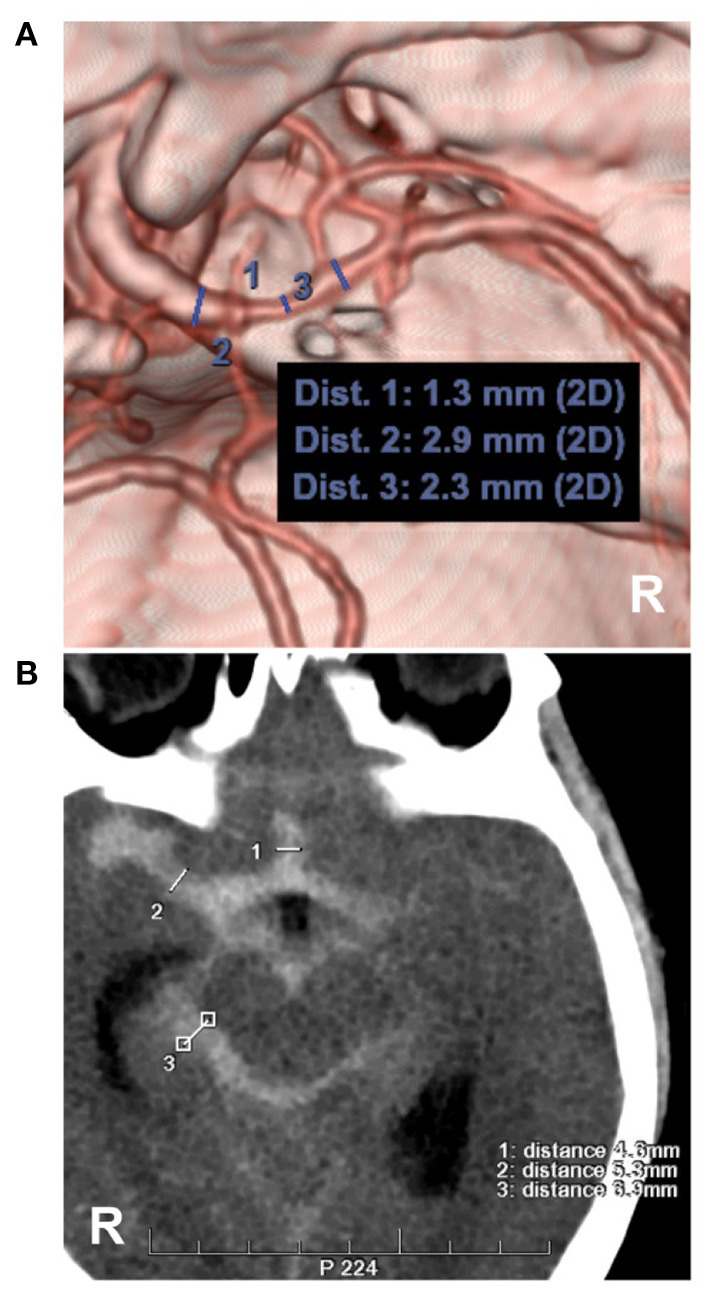
Figure 6.

Measurements of intracranial vasospasm and SAH thickness. (A) CTA showing the diameter measurement of the MCA. No. 1, 2 and 3 showing the diameters of the different locations of the MCA, the location of No. 1 had a moderate vasospasm (33–66%, 1.3/2.3 and 1.3/2.9). (B) CT showing the measurements of the Barrow Neurological Institute scale, No. 1, 2 and 3 showing the subarachnoid hemorrhage thickness (4.6, 5.3, 6.9 mm), by the measurement across the thickest-appearing regions of the cistern or fissure. CT, computed tomography; CTA, computed tomography angiography; MCA, middle cerebral artery; R, right.
