
FIGURE 2.
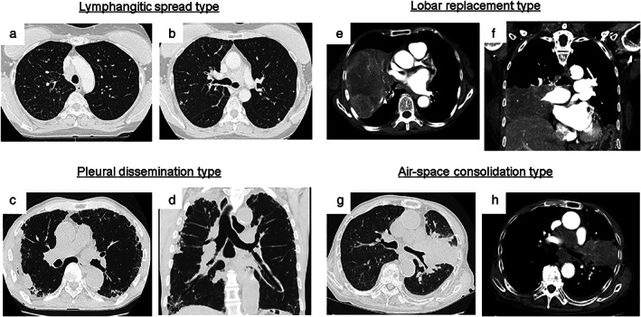
Representative images of the tumor extension and spread pattern of atypical types of small‐cell lung cancer. Typical thin‐sliced computed tomography images of the lymphangitic spread type (a and b), pleural dissemination type (c and d), lobar replacement type (e and f), and air‐space consolidation type (g and h)