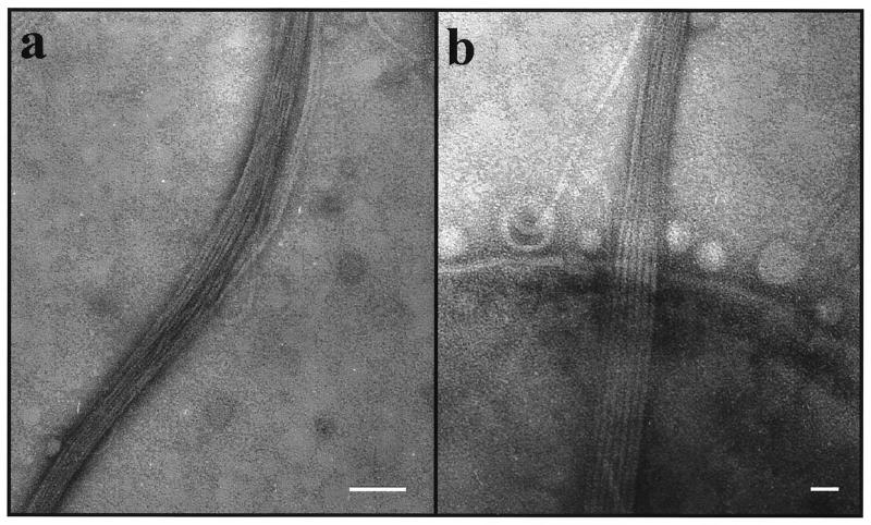
FIG. 2.
Electron micrograph of fibril bundles produced by the rough strain CU1000N. (a) Fibril bundle showing individual strands separating from the bundle at several places. Bar, 50 nm. (b) Parallel array of six or seven fibrils passing over the edge of a bacterial cell. The spherical structures at the bacterial cell surface are likely to be membranous vesicles, which are commonly seen in preparations of rough strains of A. actinomycetemcomitans (30). Bar, 20 nm.

