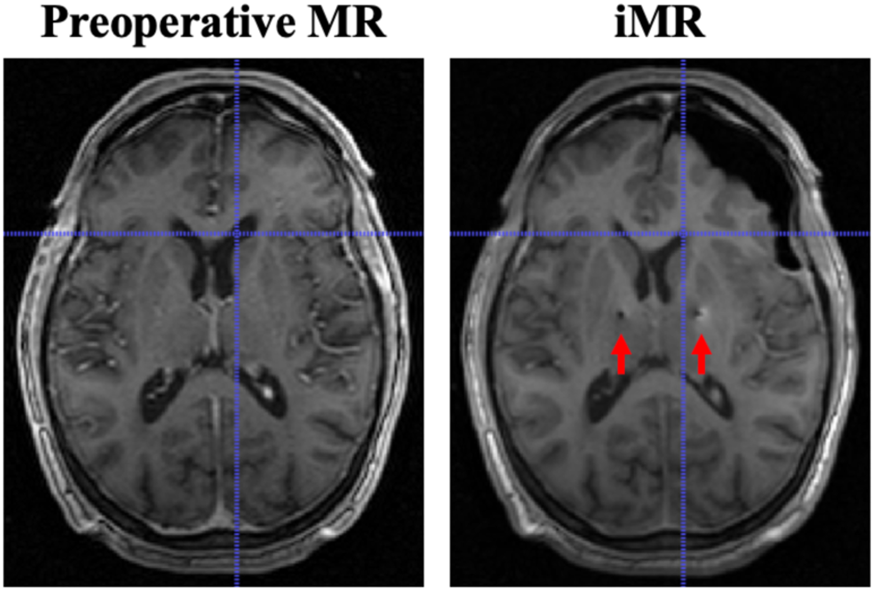
Fig. 1.

Comparison of preoperative MR and iMR imaging data on a corresponding slice. Significant asymmetric shift can be observed. Subsurface shift, e.g. at the lateral ventricle, is indicated by the crosshairs. Midline shift is also observed. The insertion path of the electrode leads and resultant imaging artifacts can be observed (red arrows) on iMR imaging data.
