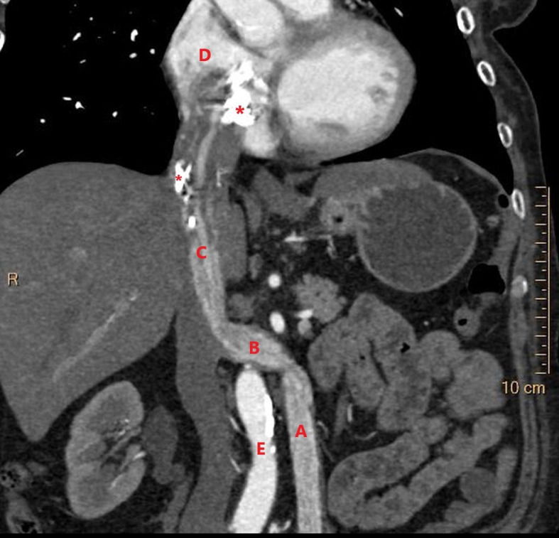
Figure 1.

Coronal reconstruction of CT scan of the thorax and abdomen in the late arterial phase. The CT image demonstrates the intravascular leiomyomatosis in the left ovarian vein (A), extending through the left renal vein (B) into the inferior vena cava (C), and progressing into the right atrium of the heart (D). (E) aorta. The upper part of the tumour is seen with several areas of calcification (*). – Courtesy of Gratien Andersen, MD.
