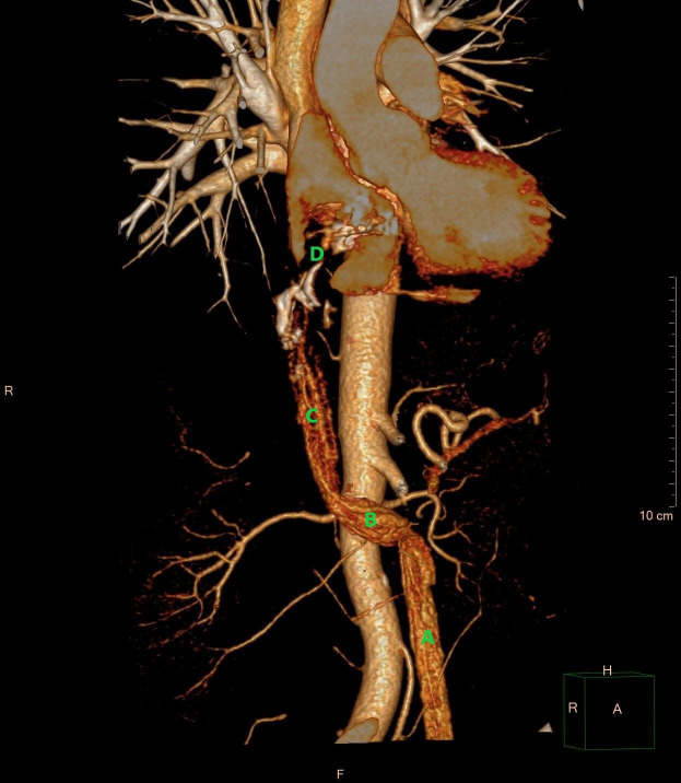
Figure 3.
3D volume rendering demonstrating an intravascular tumour in the left ovarian vein (A), the left renal vein (B), the inferior vena cava (C), and the right atrium (D). The tumour in the right atrium is shown as a contrast filling defect (D). Courtesy of Gratien Andersen, MD. 3D, three dimensions.

