Figure 5.
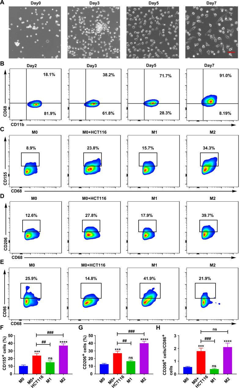
CRC cells triggered CD155 expression in human macrophages. (A) Microscopic images of differentiating macrophages from human PBMC-derived monocytes. Scale bar: 50 µm. (B) CD68 expression pattern during macrophagic differentiation of monocytes. CD155 expression (C), CD206 expression (D), and CD86 expression (E), in macrophages during co-cultured with CRC cells, M1 (LPS+IFN-γ-treated) and M2 (IL-4+IL-13-treated) polarization (gated on CD68+ cells). (F–H) Statistical analysis of CD155 expression, CD206 expression, and the ratio of CD206 to CD86 in macrophages during co-cultured with CRC cells, M1 (LPS+IFN-γ-treated) and M2 (IL-4+IL-13-treated) polarization. Data were presented as mean±SD, n=3. A significant difference between the groups, ##p<0.01 and ###p<0.001, and compared with M0 group ***p<0.001 and ****p<0.0001. CRC, colorectal cancer; IFN, interferon; IL, interleukin; LPS, lipopolysaccharide; ns, no significant difference; PBMC, peripheral blood mononuclear cell.

