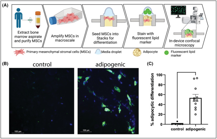
FIGURE 3.

Differentiation of primary human bone marrow mesenchymal stem cells (MSCs) in Stacks. (A) MSCs from two separate donors were seeded into Stacks and cultured for 14 days under control or adipogenic conditions. (B) Adipocytic differentiation was shown by the development of lipid droplets, identified with LipidSpot™ staining (green) and Hoechst nuclear staining (blue) by confocal microscopy. (C) LipidSpot™ ‐positive cells and total nucleated cells were quantified using automated image analysis and expressed as a percentage of LipidSpot™‐positive cells. Each data point represents percentage of LipidSpot‐positive cells in one microwell; *p < .05.
